Six of the World's Top Ten MedTech Giants Enter Intracardiac Echocardiography (ICE) Market, Signaling Industry Boom

TINGSN
Developer of Cardiac Interventional Treatment Devices

SONOSEMI
Active Vascular Implantable and Interventional Medical Device R&D and Manufacturer

ICE
Next-Generation Intracardiac Ultrasound Device Developer
Globally, well-known multinational corporations such as Johnson & Johnson, Abbott, Siemens, Philips, GE Healthcare, and Boston Scientific have all entered the intracardiac echocardiography (ICE) market.
For example, Biosense Webster under Johnson & Johnson launched the SoundStar 3D intracardiac echocardiography catheter; St. Jude Medical under Abbott introduced the ViewFlex Xtra ICE catheter; Siemens Healthineers has launched the AcuNav Volume ICE catheter; Philips has introduced the VeriSight Pro 3D ICE catheter; Boston Scientific has launched the Ultra ICE catheter...
Data shows that intracardiac echocardiography (ICE) is an innovative echocardiographic diagnostic technology. It places the ultrasound probe inside the heart chambers to emit and receive ultrasound signals, enabling real-time imaging of cardiac anatomical structures, cardiac hemodynamics, and cardiac function.
So far, intracardiac echocardiography has been applied in various surgeries such as atrial fibrillation radiofrequency ablation, left atrial appendage closure, atrial septal defect closure, mitral valve repair, and endomyocardial biopsy.
Clinical data shows that: Intracardiac echocardiography has improved the accuracy and safety of procedures such as atrial fibrillation radiofrequency ablation and left atrial appendage closure, reduced surgical risks, minimized complications, lowered procedural costs, and enhanced patient comfort and procedural efficiency. Additionally, intracardiac echocardiography can effectively reduce the use of X-rays, thereby decreasing radiation exposure for both patients and doctors.
For example, clinical studies have shown that using the AcuNav Volume ICE catheter for left atrial appendage closure can shorten the surgical process and recovery time by approximately 57% and save about 33% of potential costs.
Based on the significant clinical advantages of intracardiac echocardiography, we anticipate that it will become one of the biggest winners behind the rapid development of the cardiac electrophysiology and structural heart disease industries.
As early as 1956, researchers explored "catheter + ultrasound technology" for cardiac structure imaging. However, due to technical limitations, the exploration at that time was not successful.
It was not until 1980, after breakthroughs in ultrasound imaging technology and catheter-based intervention techniques, that intracardiac echocardiography (ICE) was first applied. However, the initial application of ICE used transducers with higher frequencies, ranging from 20-40 MHz, which had limited tissue penetration, preventing the acquisition of detailed intracardiac anatomical images. Despite this, the application of ICE during this period significantly boosted researchers' confidence and laid the foundation for subsequent development and innovation of the product.
Thereafter, researchers developed low-frequency transducers (9MHz) to enable imaging of intracardiac structures. However, these systems still have pain points such as low catheter flexibility, limited imaging depth, and the inability to fully visualize cardiac structures.
In 1990, an innovative company optimized the imaging depth of intracardiac ultrasound by using a lower frequency (5MHz). Unfortunately, the corresponding transducer was limited in clinical application due to its larger size.
With the innovative development of ultrasonic phased array technology, intracardiac ultrasound has ushered in new opportunities. The application of phased array technology in the field of intracardiac ultrasound not only addresses the issue of larger transducer size but also expands the clinical application scope of intracardiac ultrasound systems. Simply put, based on ultrasonic phased array technology, intracardiac ultrasound achieves deeper imaging depth and clearer images. At the same time, intracardiac ultrasound can provide various imaging information, such as color flow, tissue Doppler, and spectral Doppler.
In addition, the miniaturization of ultrasound transducers has also been achieved, allowing rotational intracavitary ultrasound to be widely used in clinical settings. Both rotational intracavitary ultrasound and phased-array intracavitary ultrasound have their own advantages and characteristics. For instance, rotational intracavitary ultrasound can provide higher near-field resolution, while phased-array intracavitary ultrasound can offer various information such as anatomical structures, color flow Doppler, and spectral Doppler. Currently, phased-array intracavitary ultrasound is more widely used and occupies a dominant position.
At the beginning of the 21st century, Siemens Healthineers launched the first 2D intracardiac echocardiography catheter, ACUSON AcuNav, which was initially mainly used to guide atrial septal puncture. With this intracardiac echocardiography, doctors are able to obtain atrial septal images, measure the size of atrial septal defects; determine the positional relationship between atrial septal defects and structures such as the aorta, mitral valve, tricuspid valve, superior and inferior vena cava, making procedures like atrial septal puncture more precise and safer.
Based on the product's advantages in structural heart disease, it has been subsequently expanded for use in various procedures such as atrial septal defect closure and left atrial appendage closure.
With the widespread application of intracardiac echocardiography, its performance has been gradually optimized. In the past, intracardiac echocardiography has evolved from two-dimensional imaging to three-dimensional imaging, significantly enhancing guidance and visualization capabilities. Two-dimensional intracardiac echocardiography supports dual-plane or triple-plane imaging, displaying two or three different planar views, but doctors need to mentally reconstruct these images into a three-dimensional anatomical structure. Three-dimensional intracardiac echocardiography, on the other hand, can directly present a three-dimensional anatomical structure, allowing doctors to perform surgeries more easily.
In recent years, companies like Siemens and Johnson & Johnson have innovated real-time 3D intracardiac ultrasound systems. If 2D intracardiac ultrasound is considered akin to a black-and-white television, then real-time 3D intracardiac ultrasound is comparable to a 3D IMAX movie. Compared with 2D intracardiac ultrasound, the newly introduced real-time 3D intracardiac ultrasound not only enables real-time imaging but also displays three-dimensional heart structures and blood flow information, with clearer and more precise images.
According to the product development direction, we expect that an increasing number of intracardiac ultrasound systems will evolve from two-dimensional to three-dimensional, and from three-dimensional to real-time three-dimensional imaging. Meanwhile, intracardiac ultrasound will also advance towards clearer, more precise, and multifunctional capabilities.
From the perspective of application scenarios, intracardiac echocardiography can be used in various surgical procedures such as atrial fibrillation ablation, left atrial appendage closure, atrial septal defect closure, mitral valve plasty, and endomyocardial biopsy. The targeted diseases include atrial fibrillation, congenital heart disease, cardiogenic stroke, atrial septal defect, mitral stenosis, tricuspid stenosis, and aortic stenosis.
According to the Frost & Sullivan report, in 2021, there were approximately 133,400 congenital heart disease patients, about 4.5 million cardiogenic stroke patients, around 3.9 million aortic regurgitation patients, approximately 4.4 million aortic stenosis patients, about 5.9 million mitral stenosis patients, and around 10.8 million mitral regurgitation patients in China. Meanwhile, an epidemiological survey conducted by Renmin Hospital of Wuhan University found that there are nearly 20 million atrial fibrillation patients in China. Globally, there are approximately 1.7 million congenital heart disease patients, about 19.7 million cardiogenic stroke patients, and around 220 million valvular heart disease patients.
It can be seen that the patient population associated with intracardiac echocardiography is extremely large, and the huge demand will give rise to a massive market. Currently, the market price for intracardiac echocardiography catheters is approximately 20,000 RMB per unit. With a penetration rate of 10%, the market potential for intracardiac echocardiography catheters in China will exceed 10 billion RMB. The main system for intracardiac echocardiography costs around 1 million RMB per unit. If we estimate that only one tertiary hospital in China is equipped with one main system (excluding non-tertiary hospitals), the market potential for intracardiac echocardiography main systems will surpass 1.4 billion RMB.
Although intracardiac ultrasound has broad prospects, it is still in the early stages of development.
According to the latest released "China Structural Heart Disease Industry Report 2021," in 2021, there were over 6,500 cases of transcatheter aortic valve implantation (excluding clinical research) in China, about 350 cases of transcatheter mitral valve edge-to-edge repair (TEER), approximately 14,000 cases of left atrial appendage closure, and around 75,000 cases of congenital heart disease interventional surgeries. The "China Cardiovascular Health and Disease Report 2021" shows that in 2020, the number of atrial fibrillation radiofrequency ablation procedures in China was approximately 35,000.
As of now, the penetration rate of interventional procedures for structural heart disease in China remains at a relatively low level, which also results in fewer uses of intracardiac echocardiography products for such procedures.
However, experts believe that interventional treatment for structural heart disease has not yet reached a plateau and will rapidly become widespread, increasing its penetration rate. This is because innovative devices for treating structural heart disease—such as mitral annulus repair, tricuspid annuloplasty rings, transcatheter valve replacement products, and transvenous tricuspid repair devices produced by Chinese innovative companies—have been successively approved. Moreover, these companies are stepping up efforts in market education and academic promotion.
According to the Frost & Sullivan report, the market size of interventional devices for structural heart disease in China has increased from 400 million yuan in 2017 to 2 billion yuan in 2021, with a compound annual growth rate of 48.3%. It is expected to reach 10.4 billion yuan by 2025, with a compound annual growth rate of 51%.
Based on the nearly 50% annual compound growth rate of the structural heart disease interventional device market, intracardiac echocardiography is also expected to experience rapid growth. Meanwhile, the intracardiac echocardiography market will continue to expand with the increasing adoption of atrial fibrillation ablation procedures.
With the improvement of residents' economic levels and the increasing demand for precision treatment, intracardiac ultrasound will gradually become a necessity product.
Previously, transesophageal echocardiography (TEE) and transthoracic echocardiography (TTE) were used in clinical practice. Compared with TEE, intracardiac echocardiography (ICE) is more flexible in operation and can image from inside the heart, enabling more precise detection of thrombosis and surgical guidance. TEE requires general anesthesia for special populations, while ICE only needs local anesthesia, avoiding the discomfort of esophageal intubation, as well as the mechanical damage to the esophageal mucosa caused by the TEE probe and thermal injury during scanning.
Compared with transthoracic echocardiography (TTE), intracardiac echocardiography is not affected by factors such as obesity, emphysema, and thoracic deformities, enabling shorter-distance and higher-precision imaging.
In addition, compared with traditional methods such as TEE, intracardiac echocardiography can monitor in real time the risks of potential complications during surgery, such as thrombosis and pericardial effusion, improving surgical safety. Intracardiac echocardiography is independently operated by the surgeon without relying on ultrasound or anesthesiology physicians, allowing for more flexible surgical scheduling. It also reduces personnel costs, enhances surgical efficiency, and shortens the time spent on each surgical procedure.
In clinical practice, TEE has always been the gold standard for excluding thrombosis in the left atrium and left atrial appendage before atrial fibrillation catheter ablation or left atrial appendage closure. Clinical data shows that intracardiac echocardiography (ICE) is equivalent to TEE in assessing the accuracy of left atrial appendage thrombosis, and ICE provides higher image quality. In terms of the effectiveness in diagnosing left atrial appendage thrombosis, ICE is superior to TEE.
At the same time, the intracardiac echocardiography (ICE) catheter scans layer by layer from one end of the left atrial appendage to the other, which can fully display the maximum diameter of each axis of the left atrial appendage, the landing zone diameter, and the effective working depth, thereby providing more precise guidance for occluder selection. Multicenter studies have found that ICE-guided left atrial appendage occlusion procedures take less time in each step, with higher surgical efficiency, and significantly reduced costs for anesthesia and ultrasound professionals.
In terms of atrial fibrillation, intracardiac echocardiography has long been applied in catheter ablation for atrial fibrillation. Compared with traditional X-ray imaging, intracardiac echocardiography can replace or partially replace X-ray imaging, reduce radiation exposure, and more clearly and accurately identify the intraoperative cardiac conditions.
According to a 2020 statistical report spanning 14 years and covering nearly 300,000 patients who underwent atrial fibrillation ablation, the use of intracardiac echocardiography (ICE) during catheter ablation significantly reduced in-hospital mortality. The overall complication rate decreased by 52%, and hospital stays were notably shortened (about 4 days without ICE vs. about 2 days with ICE).
Precise surgeries based on intracardiac ultrasound are low-risk, have fewer complications, require shorter hospital stays, and offer faster recovery. Therefore, we anticipate that they will be more widely applied in clinical settings.
As of now, global device giants Siemens, Philips, GE Healthcare, Johnson & Johnson, Abbott, Boston Scientific, and others have already laid out their strategies in intracardiac ultrasound. Among them, Siemens, Johnson & Johnson, and Philips have launched or developed 3D and 4D intracardiac ultrasound products.
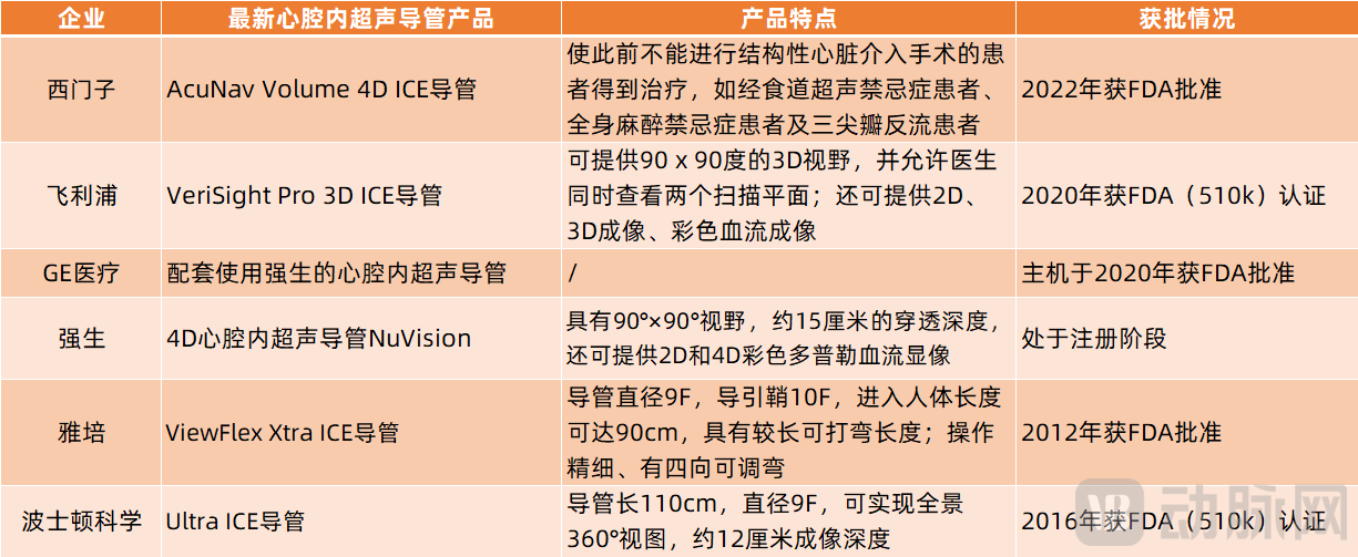
For example, Siemens' AcuNav Volume 4D ICE catheter, which received FDA approval in 2022, can provide 2D and 4D intracardiac imaging, 2D and 4D color Doppler flow imaging, as well as spectral Doppler. Clinical experts have stated that this ICE catheter expands the range of applications, allowing patients who were previously unable to undergo structural heart interventional procedures to receive treatment, such as patients with contraindications for transesophageal echocardiography, contraindications for general anesthesia, and tricuspid regurgitation.
Johnson & Johnson has developed the 4D intracardiac ultrasound catheter NuVision, which is currently in the registration application stage. Data shows that this NuVision intracardiac ultrasound catheter can perform real-time ultrasound imaging with a 90°×90° field of view, approximately 15 centimeters of penetration depth, and can also provide 2D and 4D color Doppler blood flow imaging. In addition to NuVision, Johnson & Johnson has also launched the SoundStar (3D) ultrasound catheter, which provides real-time intracardiac echocardiography imaging and navigation.
It is worth mentioning that Johnson & Johnson has collaborated with GE Healthcare. Its NuVision intracardiac ultrasound catheter is used in conjunction with GE Healthcare's Vivid Ultra Edition ultrasound system. Additionally, in 2021, Johnson & Johnson partnered with GE Healthcare to jointly develop an integrated green catheterization lab solution, leveraging their respective strengths to enable zero radiation, safer, and more precise atrial fibrillation catheter ablation procedures.
In addition, Philips has also partnered with Abbott. Abbott's ViewFlex Xtra ICE catheter can be used in conjunction with Philips' CX50 xMatrix ultrasound system.
In addition to the strategic alliances among these multinational medical device companies, innovative enterprises are also making active breakthroughs. For instance, Conavi, an innovative company incubated by a medical research institution, received FDA approval for its Foresight ICE system in 2016. In 2014, the company partnered with Japan Lifeline, a Japanese publicly listed company, which exclusively distributes Conavi’s ICE products in Japan. In 2017, Conavi collaborated with China's publicly listed company Grand Pharmaceutical, granting it exclusive rights to sell the Foresight ICE system and other products in China. With the support of publicly listed companies like Japan Lifeline and Grand Pharmaceutical, the rapid adoption of Conavi's Foresight ICE system is anticipated.
In China, there are currently four main ICE products on the market, provided by Siemens, Boston Scientific, Johnson & Johnson, and Abbott, respectively. However, innovative companies such as TINGSN, SONOSEMI, ICE, and Heart Space Technology have also begun actively developing intracardiac echocardiography, achieving breakthrough progress.
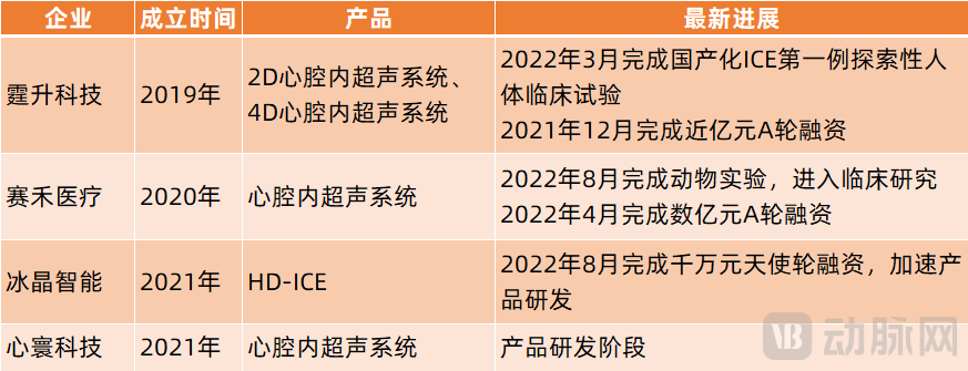
For example, TINGSN has developed 2D intracardiac echocardiography (ICE) and 4D ICE products. The former is mainly used for simple structural heart disease and electrophysiological procedures, while the latter is primarily utilized for complex structural heart disease and electrophysiological surgeries. Together, they will meet most of the needs in the structural heart disease and electrophysiology fields. In early March 2022, TINGSN successfully completed the first exploratory human clinical trial of domestically produced ICE in China, marking a breakthrough for Chinese-produced ICE.
For innovative companies like TINGSN and SONOSEMI, on the one hand, intracardiac echocardiography, structural heart disease, and atrial fibrillation ablation are still in their early stages of development with low penetration rates, indicating significant room for growth. On the other hand, the active involvement of globally renowned medical device enterprises in intracardiac echocardiography will bring immense competitive pressure to these companies.
In this regard, Zhang Dongyu, founder of TINGSN, stated: "For the industry of ICE produced in China to achieve breakthroughs, relevant companies need to focus on the development and optimization of transducer technology, connection cable technology, and packaging technology. They must ensure production efficiency and yield rate can reach mass production levels while reducing manufacturing costs as much as possible. All of these require significant investment from companies in terms of researchers, equipment, environment, funds, and time."