AstraZeneca Reveals pH-Dependent Structural Transitions in Ionizable Lipids That Enhance Endosomal mRNA Release Efficiency

AstraZeneca
Biopharmaceutical Manufacturer
Lipid Nanoparticles(LNP)Significant progress has been made in the research of rational design and screening methods, with the efficiency and safety of nucleic acid delivery having improved by several orders of magnitude through the continuous efforts of researchers. In addition to delivering small interfering RNA (siRNA),(siRNA)mRNA vaccines for drugs and infectious diseases are also expected to play an important role in cancer immunotherapy mRNA vaccines, ex vivo and in vivo engineered delivery for adoptive cell therapy, and CRISPR gene editing.
Despite the clinical success of LNP-mRNA-based therapies, the mechanism by which LNPs mediate the release of mRNA from endosomes into the cytoplasm remains incompletely understood. The circulation kinetics, biodistribution, specific targeting, and cellular uptake of LNPs can be optimized through surface property modifications, and it is generally believed that,Ionizable Lipid Components(CIL)TheStructural Transition and pH-Dependent Reaction MechanismIt is crucial for endosomal release. Currently, most studies are limited to the endosomal escape of empty LNPs, while ignoring the overall structural changes and release of mRNA and CIL in response to endosomal pH changes.
December 6, 2023, fromAstraZenecaThe R&D team in the journalPNASPublished online with the title“pH-dependent structural transitions in cationic ionizable lipid mesophases are critical for lipid nanoparticle function”Research Paper.This study systematically compared different ionizable lipids.pH-Dependent Structural Transition, and reveal through molecular dynamics simulationDifferent Effects of Structural Transition Differences on mRNA Endosomal Escape and Release。
CIL/Chol exhibits changes with varying pH.
Lyotropic Phase

To understand the structural transitions in the core of LNPs, researchers investigated the macroscopic bulk phase of CIL/chol/buffer with or without nucleic acid cargo.(Macroscopic bulk phases)The variation with pH was analyzed using high-resolution synchrotron radiation SAXS data. The monophosphate adenine homopolymer poly(A) was selected as a substitute model for mRNA due to its similar biophysical properties, and the mixtures used in the study were designed to closely simulate the preparation and production process of LNP-mRNA drugs.
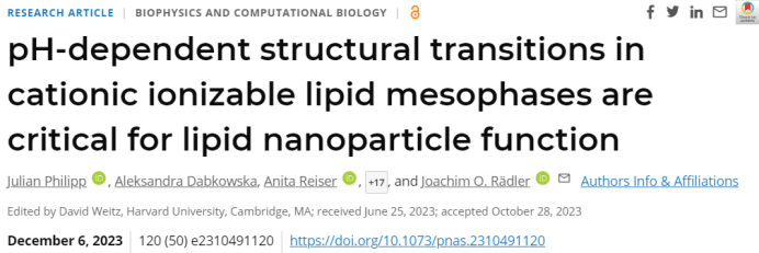
Figure 1A shows three types of CIL.(MC3, KC2, and DD)Scattering data of CIL/chol/buffer bulk phases dialyzed under a series of pH conditions, identifying the crystal symmetry of ordered phases based on relative peak positions(Figure 1C)。
The overall structural progression trend for all three CIL/chol/buffer phases is as follows:
1) InNeutral pHBelow, the main phase is the disordered inverse micellar LII phase, with a primary structural peak.(At 0.1 Å−1)And a second-order weak peak at approximately 0.2 Å−1;
2) WhenpH value is approximately 6.0 to 6.5When isotropic cubic reverse micelle phase appears(Fd3m), subsequently transforming into the reverse hexagonal HII phase.
DD/chol/buffer exhibits an additional bicontinuous cubic phase (Pn0m) at extremely low pH values below 5. In very rare cases, a P6(3)/mmc hexagonal crystal structure phase appears at pH 7. According to Figure 1A, it was also observed thatThe intermediate phase of the CIL overall sequence follows the behavior driven by the molecular shape factor during the formation of lyotropic liquid crystal phases., also known as CPP(critical packing parameter):At neutral pH, the CIL monolayer exhibits negative curvature and tends to formInverse Micelle Structure. Increase the protonation level of the CIL head group(Increases as pH decreases)This will simultaneously increase the headgroup area, subsequently causing the structural curvature to spontaneously shift towards smaller negative values, eventually approaching zero curvature.
These results indicate that the lipid bulk phases of the three CIL/chol/buffer mixtures exhibit nearly identical intermediate phase sequences.Structural trends decrease as the pH value decreases.From Negative Curvature to Zero Curvature Transition。
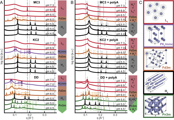
Figure 1. SAXS-based pH-dependent intermediate phase transition identification
Poly(A) Complexes with CIL in the Presence of Excess CIL

After adding poly(A) to the CIL/chol/buffer phase, an additional peak corresponding to the complex phase can be observed in the SAXS data. The additional peak appears above the phase without poly(A), thus showing the coexistence of excess CIL phase with the polyA-CIL complex phase. Two closely spaced first-order reflections are observed in the pH range of 5.5 to 6.5, confirming the coexistence of inverse hexagonal and inverse lipid phases. When the pH is above 6.5, the ordered phase transitions to a disordered LII phase, while the complexed inverse hexagonal phase continues to exist with a peak higher than LII.
During this process, no increase in nucleic acid content was found in the supernatant, indicating that poly(A) was not released from the bulk phase as the pH increased. Therefore, researchers speculated that the HcII inverse hexagonal micelles dispersed into poly(A)-filled inverse cylindrical micelles LcII and dissolved in the excess micellar lipid phase. This equilibrium phase remained stable for a long period of time, with SAXS showing no signs of multiple phase coexistence over an extended duration.
Adding poly(A) to the CIL/chol/buffer system leads to the complexation of poly(A) and CIL, resulting in an additional inverse hexagonal condensed phase HcII.。At high pH values, such as 6 to 7, a dispersed inverse cylindrical micelle LcII phase is formed.
LNP-mRNA Still Exhibits pH-Dependent Structural Change Characteristics

Next, the researchers directly compared the SAXS scattering curves of CIL+poly(A) with CIL+mRNA and LNP+mRNA at pH 5 and pH 7.(Figure 2A)。
After the mRNA replaced poly(A), no peak similar to the complex state HcII observed in the previous stage of the experiment appeared. This finding may be due toThe secondary structure of mRNA is incompatible with long-range antihexagonal order.When comparing the scattering of CIL-mRNA bulk phase with 100 nm sized mRNA-LNP, an increase in actual spatial spacing was observed. Researchers speculate that the size effect may reduce the long-range order of the inverse hexagonal phase, and the core phase composition of LNP might be slightly different from the reconstructed CIL bulk phase, but overall, they exhibit very similar scattering data. This result suggests that,In the LNP core phase, there is also a transition of CIL from the disordered LII phase at high pH to the ordered HII phase at low pH.Figure 2B shows the arrangement during the transition process. The cylindrical CIL structure will coverRetaining Secondary StructuremRNA profile, and is characterized as LcII.
Figure 2. Scattering Characteristics and pH-Induced Transitions of CIL and LNP Core Phases
pH-Induced CIL and LNP Structural Transformation and Transfection Efficiency Differences

In Figure 2C, it can be observed that MC3 and KC2 exhibit very similar trends and spacing.The nearest neighbor distance in the reverse micelle phase LII increases as the pH decreases.. This can be interpreted as the protonation of ionizable lipid head groups, accompanied by micelle hydration and swelling.As the size increases, the degree of stacking arrangement increases, eventually forming a cubic liquid crystal phase at a pH of approximately 6.5.At this pH, the coexistence of the inverse hexagonal HII phase is almost always observed. Researchers also found that,The nearest neighbor distance between MC3 and KC2 does not change with further reduction in pH.。
In contrast,DD has a larger nearest neighbor spacing.DD is characterized by the coexistence of a double-continuous cubic phase and HII.As the pH value decreases, the nearest neighbor distance gradually expands., slowly forming a cylindrical LII phase similar to MC3 and KC2 and further undergoing transformation.
Based on this, the researchers used Cy5-labeled mRNA fluorescence and eGFP expression fluorescence after LNP transfection to compare transfection efficiency and monitor the time points of endosomal fusion events.MC3 and KC2 showed similar Cy5 and eGFP signal onset times, while the onset of both Cy5 and eGFP in DD was relatively delayed. Therefore, LNPs using DD as a CIL component exhibited a lower initial expression rate of eGFP, demonstrating delayed transfection and expression kinetics.
MD simulation shows that,An excess of lipid structural components at least partially favors the formation of inverted micellar geometry and undergoes a topological transition from inverted to cylindrical shapes. This mechanism may facilitate the fusion of LNPs with endosomal membranes.
In addition, the researchers also proposed,"Endosomal Membrane Fusion"And"mRNA Release"Should be regarded as two distinct processes with a certain delay.The time difference between the two fluorescences proves,The fusion of endosomal membranes does not immediately lead to mRNA release and translation, but it is unclear whether this delay is influenced by the electrostatic interaction between mRNA and CIL.
The latest SAXS data shows,Protein adsorption-induced conformational changes can indeed affect the core structure of LNPs, and vice versa. The surface-core interrelationship implies that pH-induced phase transitions in the LNP core structure will also influence conformational changes in the surface composition.
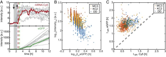
Figure 3. Expression Kinetics of eGFP-mRNA LNP Transfection In Vitro
Summary

Although most literature attempts to derive structure-activity relationships, this article demonstrates that structural phase transitions may also impact transfection activity. Similar pH-dependent structural transitions might also exist in other optimized ionizable lipids used for mRNA delivery, such as ALC-0315 and SM-102, but more in-depth comparative studies are still needed.
Studying the influencing factors of CIL structural transformation, molecular dynamics, and the effects on endosomal membrane fusion and nucleic acid cargo release will help in the rational design of mRNA-LNP formulations, enabling their future use in a broader range of medical applications.
References
R, Brummer C, et.al., pH-dependent structural transitions in cationic ionizable lipid mesophases are critical for lipid nanoparticle function. Proc Natl Acad Sci U S A. 2023 Dec 12;120(50):e2310491120. doi: 10.1073/pnas.2310491120.
E.N.D

Previous article recommendations:
Gene Therapy Approved by FDA for Market Launch, Bluebird Bio Plummets 40%
Ru Jian Pharmaceutical's First iPSC Drug Approved for Clinical Trials
【Bookmark】In-depth Industry Report: Innovative Vaccine and Nucleic Acid Vaccine Technologies
The Core Technology Behind the First CRISPR Gene-Editing Therapy Priced at $15.7 Million
LianBio Rejects Acquisition Offer
Genscript Bio CD20 NK Cell Therapy IND Accepted for Review
Research Report | CAR-T: Bringing a Ray of Hope for a Cure to Systemic Lupus Erythematosus
Review and Market Launch of Small Nucleic Acid Drugs
Rongcan Bio Completes Over 100 Million Yuan in Series A Financing
CXO Leader Reveals Harsh Industry Truth, Triggering Sector Plunge
CARsgen Pharma's GPRC5D CAR-T Therapy IND Approved by FDA
CRISPR/Cas9 Gene-Edited TIL Cell Therapy Approved for Clinical Use
$32,000/Bottle! BioMarin's Hemophilia AAV Gene Therapy Launches in Germany
Shanghai Pharmaceutical Group's Oncolytic Virus Therapy Approved for IND
Quality Control of CAR-T Cells: Ensuring Safety, Efficacy, and Stability
Hengsai Biotech Completes Hundred Million-Level Series A Financing
Disclaimer: Source RNAScript, compiled by Vergil. This article aims for knowledge sharing. All content is for reference only and does not constitute any advice.