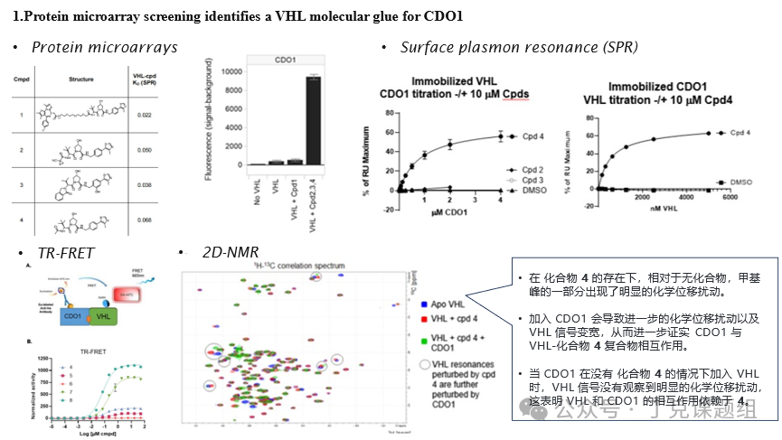
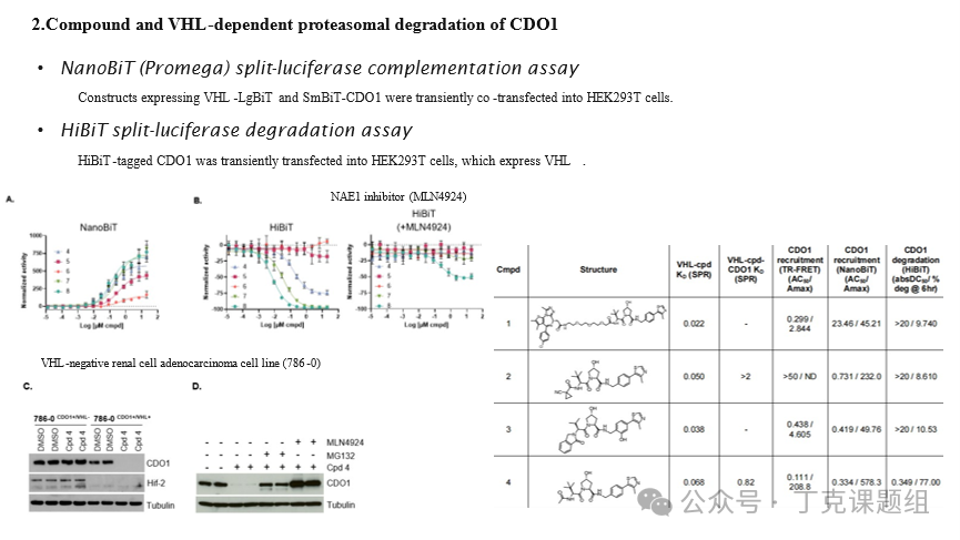
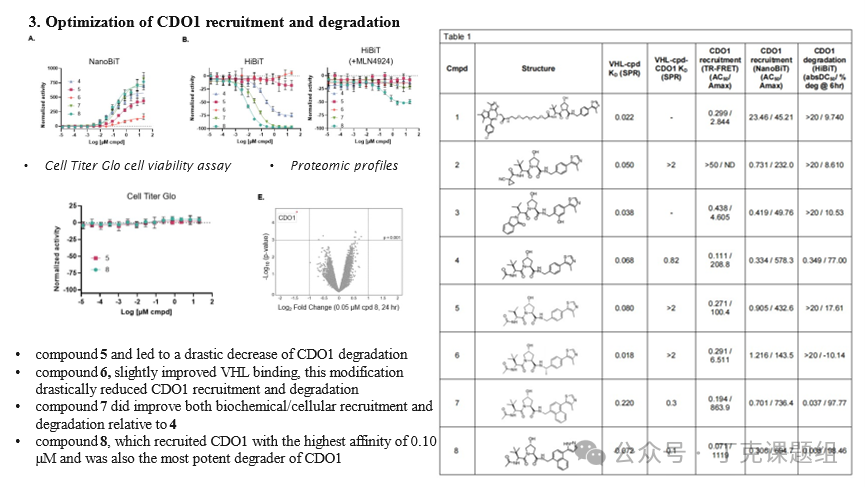
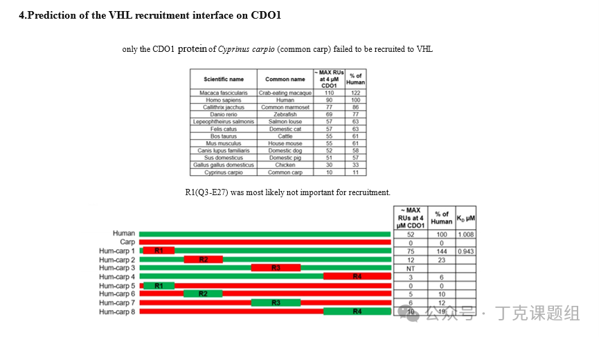
In recent years, molecular glues as a new class of chemical probes and a novel paradigm for targeted protein degradation drug development have made significant progress.2014Year,ScienceJournalReported that lenalidomide can induce CRBN Degradation IKZF1/3, this discovery has greatly promoted the development based onCRBNAdvances in the Research of Molecular Glue Degraders for Ligands. Several thalidomide-class molecules have entered clinical research stages.CRBNLigand withVHLLigands are allPROTACThe most commonly used in molecular design and synthesisE3Ligase ligand. However, so far there is no information aboutVHLThe high synergy small molecule gel degrader was reported. Recently, Novartis released on a preprint server.bioRxivPublished an article titled“A small molecule
VHL molecular glue degrader for cysteine dioxygenase 1”An article introducing a targetedCDO1TheVHLMolecular glue, it can promote VHL-Ligand-CDO1 Assembly of the ternary complex, thereby achieving VHL Dependence Degradation Endogenous CDO1。Based on Protein ChipTechnical Screening and IdentificationVHL Molecular Glue Targeting CDO1To screen and identify new substrates that VHL ligands can recruit, Novartis utilized high-content functional protein microarray (ProtoArray®) technology. These microarrays contain over 9,000 purified human proteins. Detection was performed on the bio-VBC group, DMSO group, Compound 1 group, and a mixture of three previously published VHL-binding molecules (Compounds 2, 3, and 4). Although Compounds 1-4 all bind to pVHL with similar affinity, the results showed strong signals corresponding to cysteine dioxygenase 1 (CDO1) only in the mixture group (containing Compounds 2, 3, and 4), while no signal was observed in the Compound 1 group. This suggests that one or more compounds in the mixture group are components inducing the bioVBC-CDO1 interaction. To verify this, full-length human CDO1 was expressed and purified from E. coli, and surface plasmon resonance (SPR) was used to test compound-induced interactions with bioVBC, where bioVBC was immobilized on streptavidin chips, and purified CDO1 was titrated on the surface in the presence of Compounds 2, 3, or 4. No interaction between CDO1 and bioVBC was observed when titrating 4 µM CDO1. In contrast, after adding 10 µM Compound 4 to CDO1, the binding between CDO1 and bioVBC reached saturation, with a calculated KD of approximately 0.82 µM. Additionally, neither 10 µM Compound 2 nor 10 µM Compound 3 led to a robust interaction between CDO1 and bioVBC. To confirm these results, TR-FRET and 2D-NMR were also used to verify that Compound 4 could induce the formation of a complex between pVHL and CDO1.Although these results suggest that CDO1 is recruited to pVHL via compound 4, they do not exclude the possibility that compound 4 independently binds to both CDO1 and pVHL. To investigate whether compound 4 has affinity for CDO1 in the absence of pVHL, the SPR experimental setup was reversed, with biotinylated CDO1 (bioCDO1) immobilized on a streptavidin chip. In this SPR configuration, no interaction was observed between bioCDO1 and compound 4 at titration concentrations up to 10 µM in the absence of pVHL (data not shown), nor was any interaction observed between bioCDO1 and non-biotinylated VBC, consistent with NMR observations. In contrast, when non-biotinylated VBC and compound 4 were added simultaneously, strong interaction between VBC and immobilized bioCDO1 was observed, with a calculated KD of approximately 0.49 µM.Figure 1-Screening and Identification of CDO1-VHL Molecular GlueCDO1Compounds andVHLDependent Proteasome DegradationCDO1 expression is restricted to certain tissues, primarily the lung, duodenum, small intestine, liver, and appendix, making it easily overlooked in proteomic screenings. To test whether compound 4 could recruit CDO1 to pVHL in cells, the authors used the NanoBiT (Promega) split-luciferase complementation assay for validation. Eukaryotic expression vectors expressing VHL-LgBiT and SmBiT-CDO1 were transiently co-transfected into HEK293T cells. In the presence of compound 4, a strong dose-dependent ternary complex formation signal was observed, with an AC50 of ~334 nM and an Amax of 578. Consistent with extracellular testing results, compound 3 did not form the VHL-CPD-CDO1 ternary complex, while compound 2 showed moderate recruitment activity, with an AC50 of ~731 nM and an Amax of 232.Next, the author used the HiBiT split luciferase degradation assay to verify whether the recruitment of VHL by CDO1 in the presence of the compound would lead to the degradation of CDO1 in cells. HiBiT-tagged CDO1 was transiently introduced into HEK293T cells expressing VHL and treated with different concentrations of compound 4 for 19 hours. The results showed that compound 4 could induce CDO1 degradation (absDC50 of 349 nM, 77% degradation efficiency), while compounds 2 or 3 showed no degradation activity. Subsequently, the author used a VHL-negative renal cell adenocarcinoma cell line (786-0) to validate whether the degradation results were VHL-dependent. The CDO1 gene was transduced into 786-0 cells via a viral vector (endogenous CDO1 was undetectable in 786-0 cells by Western blot). The 786-0 cells expressing CDO1 were then transfected with either a wild-type VHL vector or an empty vector to generate 786-0CDO1+/VHL+ and 786-0CDO1+/VHL- cell lines, respectively. As expected, the VHL-regulated endogenous HIF complex subunit HIF-2 was degraded in 786-0CDO1+/VHL+ cells but not in 786-0CDO1+/VHL- cells. After treatment with 0 µM of compound 4 for 16 hours, CDO1 degradation was observed in 786-0CDO1+/VHL+ cells but not in 786-0CDO1+/VHL- cells. The degradation of CDO1 induced by compound 4 in 786-0CDO1+/VHL+ cells was blocked in the presence of 1 µM MLN4924 or 1 µM proteasome inhibitor MG132.Figure 2-CDO1-VHL molecular glue can induce the degradation of CDO1 in cellsOptimize the chemical structure of VHL molecular glue to enhance the recruitment and degradation activity of CDO1To investigate the structure-activity relationship of compound 4 as a molecular glue for VHL and CDO1, the authors synthesized several analogs. Replacing the tert-butyl group on the left side of compound 4 with an isopropyl group yielded compound 5, which led to a sharp decrease in CDO1 degradation to 18%, correlating with reduced recruitment of CDO1 as observed in TR-FRET, SPR, and NanoBiT assays. Given that compound 5 exhibits similar binding affinity to pVHL as compound 4, this suggests that the additional methyl group may be important for CDO1 recruitment. Studies on the right side showed that while adding a methyl group near the benzylic position to generate compound 6 slightly improved binding to pVHL, this modification significantly reduced CDO1 recruitment and degradation. Compared to compound 4, converting the phenyl ring to a larger naphthyl ring resulted in compound 7, which demonstrated enhanced recruitment and degradation activity in both biochemical and cellular assays. Finally, replacing the methylthiazole ring of compound 7 with a pyrazole yielded compound 8, which recruits CDO1 with the highest affinity of 0.10 μM and is also the most potent degrader of CDO1 (absDC50 of 8 nM, 98% degradation). For all compounds degrading CDO1, MLN4924 was able to block or strongly reduce their degradation. To rule out the possibility that these VHL ligands cause cell death, the authors performed CellTiter-Glo cell viability assays using compounds 5 and 8 (representing very weak and potent degraders, respectively). Neither ligand had any detectable effect on cell viability.In order to explore compounds8The selectivity, the author uses 50 nM Compound8Processing Huh-7 Cell24 hours, and assess the proteomic profile using mass spectrometry.In9478In a quantitative protein, using a compound8Processing observable endogenousCDO1The robust and selective degradation, withDMSOCompared with the control group, the change is greater than3.5Times,pValue less than1.0E-003, with an overall degradation rate of62%。In the biological triple sample,CDO1Four unique quantitative peptides were identified, confirming their significant regulatory effects.Figure 3-SAR Study of CDO1-VHL Molecular GluePrediction CDO1 AndVHLContact InterfaceThe author used computer-aided protein-protein docking to simulate the potential VHL-4-CDO1 ternary complex. In the first docking step, 17 protein-protein complexes mediated by Compound 4 were generated. The complexes were classified based on the position of the ligand on CDO1, resulting in six possible ligand-binding regions of CDO1. Local protein-protein docking was performed around these six regions, and poses were scored and clustered to obtain eight possible complexes. Molecular dynamics (MD) simulations and WATMD2 water solvation analysis were further applied to evaluate the complexes and determine the water structure at the protein-protein interaction (PPI) interface. Based on the degree of dewetting observed in the calculated water structures at the PPI, a highly reliable contact interface was identified.At the same time, the author used CDO1 mutants from various species to determine the key regions at the interface. To this end, the author expressed and purified CDO1 proteins from 11 species, then tested the formation of ternary complexes in the presence of compound 4 using SPR for each CDO1 variant. The results showed that only the CDO1 protein from carp failed to be recruited to VHL. Sequence alignment of human and carp CDO1 revealed regions with sequence differences, which we randomly divided into four regions, labeled as: R1(Q3E27), R2(A35-Q65), R3(N90-R126), and R4(I145-L197). By swapping the four corresponding regions between human and carp CDO1, eight hybrid human-carp CDO1 proteins were designed, expressed, and purified, and their ability to form ternary complexes was tested by SPR. Swapping carp R1 into human CDO1 (hum-carp 1) did not result in recruitment, while swapping human R1 into carp CDO1 (hum-carp 5) did not restore recruitment. These data suggest that R1(Q3-E27) is likely not important for recruitment. Swapping the R2, R3, or R4 regions in other hybrids was associated with reduced recruitment of CDO1, suggesting that R2(A35-Q65), R3(N90-R126), and R4(I145-L197) may be important for recruitment.Figure 4 - Validation of the VHL-CDO1 interface through human-carp CDO1 protein sequence swappingThen, six narrower regions were further divided within the initial R2, R3, and R4 areas, with refined region annotations as R2'(D43-A48), R3''(M97-N101), R3'''(S114-E122), R3''''(R126), R4''(P150), and R4'''(D162-N200). Similarly, exchanges were conducted between the human and carp CDO1 protein sequences, resulting in 12 human-carp hybrid proteins, which were tested for compound 4-mediated VHL recruitment using SPR. Analysis of the results indicated that the human R2'(D43-A48) and R4'(P150) play a crucial role in CDO1 recruitment. The possible recruitment interface identified through protein-protein docking is consistent with the experimentally determined recruitment-required regions.Figure 5- Further subdivision of the region to validate the VHL-CDO1 contact interfaceVHL-cpd-CDO1 Ternary Complex X Radiation StructureThe author also successfully obtained the X-ray crystal structures of the ternary complexes VHL-cpd4-CDO1 and VHL-cpd8-CDO1. Both compounds form hydrogen bond interactions with the side chains of VHL residues Y98/R107/H110/S111/H115, while new VHL-CDO1 PPI interactions are present around the binding site. Multiple protein-protein interactions stabilize the interface, involving 12 and 14 residues from VHL and CDO1, respectively, effectively shielding compounds 4 and 8 from the solvent.CDO1's Q99 forms two hydrogen bonds with both compounds 4 and 8. Q99 is conserved in both human and carp CDO1 sequences. To validate the hypothesis that CDO1 Q99 is making critical H-bond contacts, Q99 was mutated to asparagine (Q99N), and its recruitment was tested via SPR. Compared to wild-type CDO1, CDO1(Q99N) showed over 40-fold reduced recruitment of compound 4-mediated VHL, with KD >40 μM, and a 47-fold reduction in compound 8-mediated recruitment, with KD approximately 6.1 μM. TR-FRET results were similar, showing that CDO1(Q99N) had about a 158-fold reduction in AC50 under compound 4, with AC50 around 17.4 μM, and approximately a 29-fold reduction in AC50 under compound 8, with AC50 approximately 0.87 μM. Transiently expressed HiBiT-tagged CDO1(Q99N) did not exhibit significant degradation in the presence of either compound 4 or 8. Taken together, these results indicate that CDO1(Q99) has critical interactions with both compounds 4 and 8, which are essential for robust recruitment and degradation in cells.The X-ray crystal structure also shows that the tert-butyl group on the left side of the molecule fits perfectly into the CDO1 hydrophobic patch, which is formed by the residues P44, W47, A48, A51, and V152. The naphthalene ring on the right side of compound 8 aligns with the pocket formed by residues F53, D54, Q55, G78, and P150. The naphthalene ring fills the hydrophobic cavity better relative to the benzene ring, and there is also a water-mediated hydrogen bond interaction between compound 8 and Q55.Figure 6 - X-ray structure of the VHL-cpd-CDO1 ternary complexIn summary, Novartis reported a molecular glue for VHL that promotes the formation of the VHL-cpd-CDO1 ternary complex, thereby achieving VHL-dependent degradation of endogenous CDO1. The X-ray crystal structure of the ternary complex reveals key interactions driving the recruitment of CDO1 to a novel protein interaction surface created on VHL by the degrader.In addition, CDO1 is an enzyme that breaks down cysteine by adding an oxygen molecule to the sulfur of cysteine, forming cysteine sulfinic acid. In the liver, the function of CDO1 is to eliminate excess cysteine obtained from the diet, as high levels of cysteine are cytotoxic to neurons and cells. In the article, Novartis admitted that they are not sure which diseases might benefit from CDO1 degradation. However, they have expanded the application of VHL molecular glue to other targets, with a chemical structure different from the VHL molecular glue discussed in this article. Novartis' findings allow us to explore novel VHL molecular glue for targeting proteins or classes of proteins that currently cannot be degraded using CRBN IMiDs or bifunctional molecular drugs. Furthermore, it raises the question of whether all E3 ligase ligands can be modified to become a new type of molecular glue, potentially opening up a new avenue for the screening and development of molecular glue. References:(1) Kronke, J. et
al. Lenalidomide causes selective degradation of IKZF1 and IKZF3 in
multiple myeloma cells. Science 343, 301-5 (2014).(2) Matyskiela, M.E.et al. A novel cereblon modulator recruits GSPT1 to the CRL4(CRBN)
ubiquitinligase. Nature 535, 252-7 (2016).(3) Buckley, D.et al.A
small molecule VHL molecular glue degrader for cysteine dioxygenase 1,biorxiv, doi: https://doi.org/10.1101/2024.01.25.576086