Peking Union Medical College Hospital and Tuopai Medical Launch Ultra-Widefield OCT with Curvature Analysis for Myopic Retinoschisis Assessment
TowardPi
High-end Ophthalmic Medical Device Developer

Assessment of the Severity and Related Factors of Macular Retinoschisis (MRS) and Paravascular Retinal Schisis (PVRS) in High Myopia (HM) Using UWF SS-OCT and a Novel Gaussian Curvature (K).
Data Collection:From January to November 2022, a total of 55 patients (108 eyes) with high myopia meeting the requirements were recruited from the Department of Ophthalmology at Peking Union Medical College Hospital (PUMCH).
Sample Selection:Selection criteria: High myopia (spherical equivalent greater than -6 diopters or axial length greater than or equal to 26.5 mm), age greater than or equal to 18 years, no history of vitreoretinal surgery, and no other ocular complications. Exclusion criteria include: age less than 18 years, intraocular pressure exceeding 21 mmHg, history of vitreoretinal surgery, other ocular complications such as glaucoma, keratoconus, uveitis, age-related macular degeneration or other retinal vascular diseases, cataracts or nystagmus leading to poor image quality, and incomplete medical records.
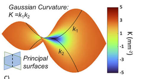
UWF SS-OCT and en face Image Acquisition and Analysis: All UWF SS-OCT images were acquired using the TowardPi Beiming BM-400K.The device adopts SS-OCT with a wavelength of 1060nm, a scanning speed of 400,000 A-Scan/second, a lateral resolution of 10μm, and an axial optical resolution of 3.8μm, with an A-Scan depth of 6.0mm (2560 pixels). Twelve radial scans were performed around the macular center, along with a 3D volume scan of 24x20mm centered on the macula (each scan containing 1280 B-Scans, each B-Scan containing 1536 A-Scans, i.e., the spacing between A-Scan and B-Scan is 15.625μm), providing a total field of view of up to 120° for subsequent enface image reconstruction. The Gaussian curvature (K) is calculated by the instrument's built-in software; the larger the absolute value of K, the steeper the local retinal shape. When K is positive, it indicates a concave retinal shape, and when K is negative, it indicates a convex retinal shape.
2. Image Analysis: Quantify local retinal shape on 3D retina using Gaussian curvature (K) and automatically identify the locations of the three highest K values (K1, K2, K3). Measure the highest K values in various regions of the retina, including the macular area (Kmax-m), superonasal vascular area (Kmax-sn), superotemporal vascular area (Kmax-st), inferonasal vascular area (Kmax-in), and inferotemporal vascular area (Kmax-it).
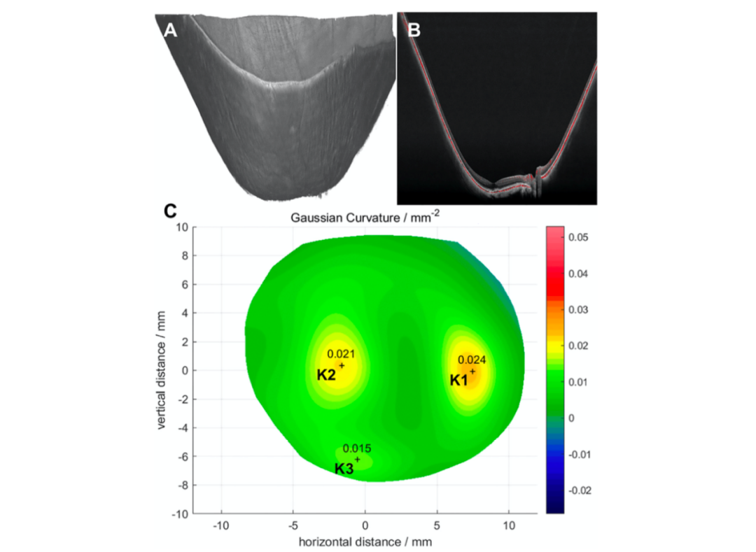
Figure 1. Typical diagram of Gaussian curvature (K) measurement. A. 3D image of the posterior segment of the eye. B. UWF-OCT scan through the macular fovea. C. Gaussian curvature topography of HM eyes, with the three highest K values marked (K1, K2, K3). HM=high myopia; UWF-OCT=ultra-widefield OCT.
3. The shape classification of posterior scleral staphyloma conforms to the criteria proposed by Ohno-Matsui et al.
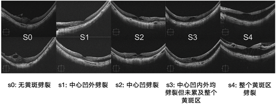
4. MRS Severity Classification: Based on the location and size of MRS, it is divided into sparing the fovea (s0), outside the fovea (s1), only the fovea (s2), involving the fovea but not the entire fovea (s3), and the entire fovea (s4).
5. The severity assessment method for PVRS is as follows: Divide the 24×20mm en face image into four quadrants (superonasal, superotemporal, inferonasal, and inferotemporal) to evaluate the presence of PVRS (its presence is also confirmed in 12 radial UWF SS-OCT B-scan images). Based on the presence and extent of PVRS, each quadrant is assigned a score from 0 to 2 (0 indicates absence, 1 indicates PVRS area <1 disc area, and 2 indicates PVRS area ≥1 disc area). The scores from the four quadrants are summed to obtain the total PVRS score for the entire 24×20mm area of that eye.
6. The severity classification of retinoschisis is revised according to the ATN classification criteria for myopic maculopathy proposed by Ruiz-Medrano et al.: Based on 12-line radial scans within a 24×20mm range for each eye, it is divided into six stages. These include: no retinoschisis (S0), inner or outer retinoschisis (S1), both inner and outer retinoschisis (S2), localized posterior pole or peripheral retinal detachment (S3), full-thickness or peripheral retinal tears (S4), and diffuse retinal detachment (S5).
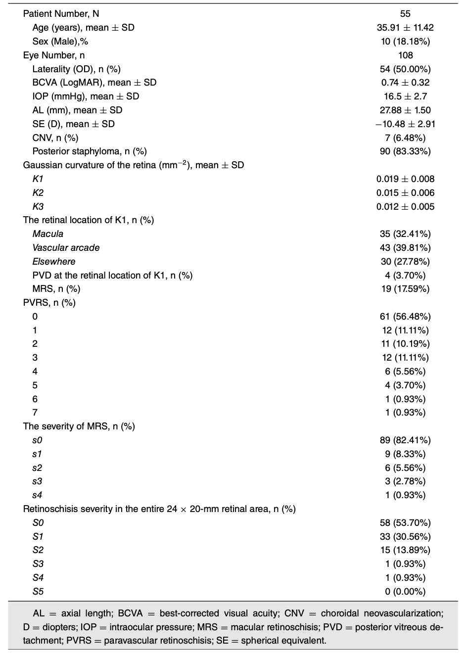
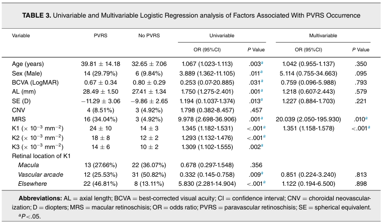
The average values of K1, K2, and K3 were ± 0.008, 0.015 ± 0.006, and 0.012 ± 0.005 mm−2, respectively. K1 was predominantly located at the vascular arcade (43, 39.81%), followed by the macular region (35, 32.41%) and other retinal areas (30, 27.78%). PVD was detected at the K1 location in 4 eyes (3.70%) included in the study but was not observed in eyes with MRS. Posterior scleral staphyloma was observed in 90 eyes (83.33%), and there was a statistically significant association between the shape classification of posterior scleral staphyloma and the retinal location of K1 (P = .002) (Table 1). MRS was detected in 19 eyes (17.59%): extrafoveal MRS (s1) in 9 eyes (8.33%), fovea-only MRS (s2) in 6 cases (5.56%), fovea but not entire macular MRS (s3) in 3 cases (2.78%), and entire macular MRS (s4) in 1 case (0.93%). PVRS was present in 47 eyes (43.52%) (Figures 2 and 3). Regarding the severity of retinoschisis across the entire 24×20mm retinal range, no retinoschisis (S0) was found in 58 eyes (53.70%), both inner and outer retinoschisis (S1) in 33 eyes (30.56%), coexisting inner and outer retinoschisis (S2) in 15 eyes (13.89%), localized posterior pole or peripheral retinal detachment (S3), and full-thickness or peripheral retinoschisis (S4) in 1 eye (0.93%). No eyes exhibited diffuse retinal detachment (S5) (Table 1).
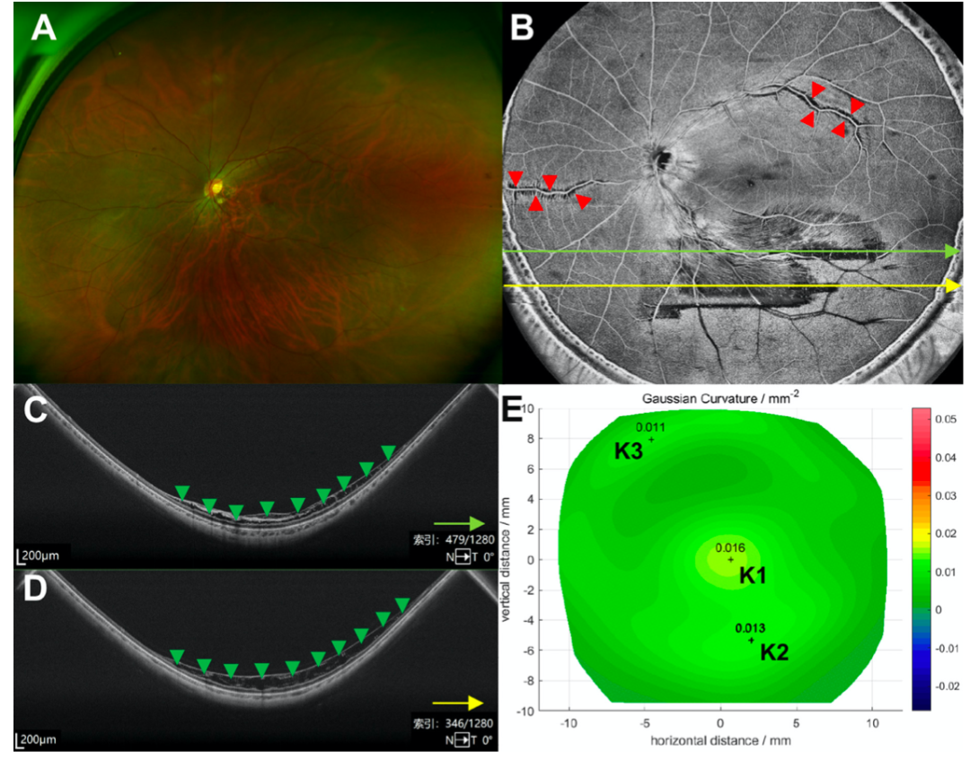
Figure 2. Typical images of an HM patient with MRS (inner retinoschisis) and PVRS. A. UWF fundus photograph. B. Corresponding 24 × 20 mm enface image. PVRS appears as feathery or streak-like hyporeflective areas along the contour of the retinal vascular arcade (red arrowheads). C and D. OCT marked with green and yellow arrow lines, corresponding to the same arrow-marked scan lines in Figure B, showing inner (green arrowhead) and outer retinoschisis (yellow arrowhead). E. Gaussian curvature (K) map. In this eye, K1 and K2 correspond to Kmax-m and Kmax-it, respectively. HM = high myopia; MRS = macular retinoschisis; PVRS = paravascular retinoschisis; UWF = ultra-widefield; UWF-OCT = ultra-widefield optical coherence tomography.
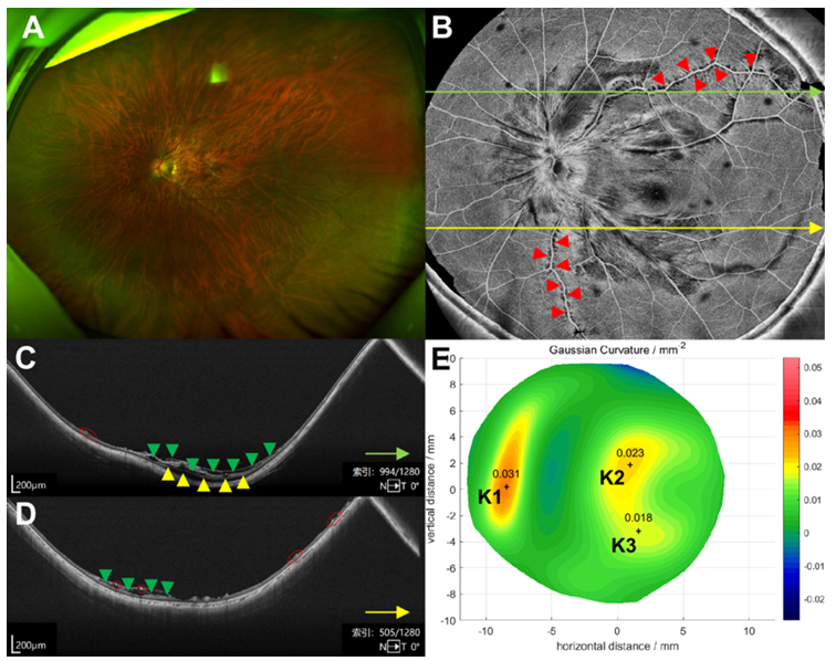
Fig. 3. Typical images of an HM patient with MRS (inner and outer retinoschisis) and PVRS. A. UWF fundus photograph. B. Corresponding 24 × 20 mm en face image. PVRS appears as feathery or streak-like hyporeflective areas along the contour of the retinal vascular arcade (red arrowheads). C and D. OCT images marked with green and yellow arrow lines, corresponding to the same arrow-marked scan lines in Fig. B, showing inner (green arrowhead) and outer retinoschisis (yellow arrowhead), as well as PVRS (red dashed circle). E. Gaussian curvature (K) map. In this eye, K2 and K3 correspond to Kmax-st and Kmax-it, respectively. HM = high myopia; PVRS = paravascular retinoschisis; UWF = ultra-widefield; UWF optical coherence tomography.
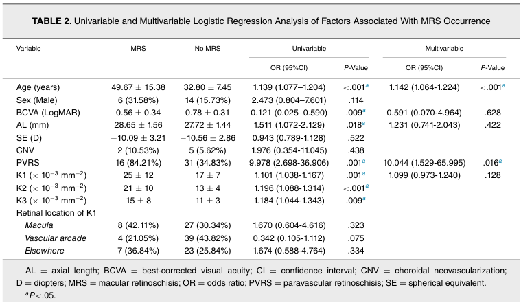
MRSRisk Factors: Univariate logistic regression analysis showed that age and PVRS were significant risk factors for the occurrence of MRS (P < 0.05). Multivariate logistic regression analysis further confirmed the correlation between age, PVRS, and the occurrence of MRS (Table 2).
Conclusion
This study shows that,Large Gaussian curvature is associated with the presence of MRS and PVRS, as well as the severity of retinoschisis. UWF-SS-OCT can visualize both central and peripheral retinal areas and is expected to become an imaging technology for the early detection of peripheral retinoschisis.Future research can further validate these findings and explore factors associated with the severity of MRS.
Discussion
This study is the first to apply ultra-widefield swept-source optical coherence tomography (UWF-SS-OCT) to evaluate the severity of retinoschisis within a large field of view, extending beyond the macular region. The study foundThe highest Gaussian curvature (K1) is mainly located in the vascular arch area, followed by the macular region and other retinal areas.The location of K1 is related to the shape classification of posterior scleral staphyloma. The study also found that more than half of the highly myopic (HM) eyes did not have retinoschisis (S0), while S1 and S2 accounted for 30.56% and 13.89%, respectively. Multivariate logistic regression analysis confirmed that age and PVRS are significant risk factors for the occurrence of MRS in HM patients.

Congratulations again to Peking Union Medical College Hospital for their research achievements in retinoschisis associated with high myopia. We look forward to the "TowardPi Retinal Morphology Triad" function, jointly developed, bringing greater well-being to patients with high myopia!


M.D., Associate Chief Physician, Associate Professor, Master's Graduate Supervisor. Deputy Director of the Department of Ophthalmology at Peking Union Medical College Hospital. Visiting Scholar at the Massachusetts Eye and Ear Infirmary (MEEI), affiliated with Harvard Medical School, USA. Participated six times in the foreign aid "Light Journey" medical team. Principal investigator for key national research projects and special clinical research funds from high-level central hospitals, published over 20 articles in Chinese and English, obtained 4 national patents, and co-authored 9 professional books. Council member of the Chinese Society of Microcirculation, committee member of the Ocular Microcirculation Professional Committee, committee member and secretary of the "Belt and Road" Ophthalmology Alliance, committee member of the Beijing Ophthalmology Residency Standardized Training Professional Committee, and youth committee member of the Beijing Medical Association Ophthalmology Branch.

TowardPi Reading Series:
Answer Revealed! Can Ultra-Widefield OCTA Replace Fluorescein Angiography for Diabetic Retinopathy?
Not Just Replacement, but Surpassing! Wide-Angle Swept-Source OCTA Replaces FFA for RVO!
New Evidence! Ultra-Widefield OCTA Can Replace Traditional Angiography in Diabetic Retinopathy
Massive Subretinal Hemorrhagic PED Secondary to PCV: Rapid Postoperative Resolution, AJO Report
Advancements in Swept-Source OCT and Swept-Source OCTA (English Translation)
"Dolphin" Leaping Out of the Retina — A Case of Congenital Vitreous Cyst


WeChat Official Account
WeChat Video Channel

Every like and view you give, I sincerely take as a sign of affection.