Organoid-on-a-Chip Platform Accelerates Mechanistic Insights into GUCY2C-Targeted CAR-T Cell Therapy for Solid Tumors

Imunopharm
Developer of Gene and Cell Therapy Technologies

At the forefront of modern medical research, organoid technology is gradually emerging, bringing new hope to cancer research.On December 18, 2024, the Chinese Academy of Engineering released the "Global Engineering Frontiers 2024" report in Beijing. The report emphasized key content in relevant fields, among which the application of tumor organoid chips in drug screening in the medical and health field has received significant attention.1Organoids, as a model capable of simulating the structure and function of human organs in vitro, provide a novel platform for an in-depth understanding of tumor development mechanisms, drug screening, and treatment efficacy evaluation.In the field of cancer treatment, chimeric antigen receptor T-cell (CAR-T) therapy has emerged as a highly promising approach, particularly demonstrating significant success in the treatment of hematological malignancies. However, its application in solid tumor treatment still faces numerous challenges, such as the lack of tumor-specific antigens, heterogeneity in antigen expression, and insufficient T-cell persistence. Guanylate cyclase C (GUCY2C) has been identified as a suitable gastrointestinal tumor antigen for developing targeted therapies, including bispecific antibodies and CAR-T. Nevertheless, there is currently no in-depth research on the optimal GUCY2C CAR-T design targeting human solid tumors and its underlying mechanisms.
1 Chinese Academy of Engineering Releases 2024 Global Engineering Frontiers in Beijing|Engineering
In September 2024, a research team led by Shen Lin from Peking University Cancer Hospital, in collaboration with Beijing Yimiaoshenzhou Medical Techology Co., Ltd., published a study titled "Antigen independent activation is critical for the durable antitumor effect of GUCY2C-targeted CAR-T cells" in the *Journal for Immuno Therapy of Cancer* (IF: 10.9). The study utilized organoid models to comprehensively investigate the efficacy and potential of GUCY2C-targeted CAR-T cell therapy in antitumor treatment, aiming to pave new paths for cancer therapy and offer more innovative and efficient treatment strategies.
01
Research Methods
● Adopting multiple experimental methods:Including cell culture and transduction, sorting to obtain different GUCY2C-expressing cells and related green fluorescent protein (GFP) cells, performing affinity detection of anti-GUCY2C single-chain antibody fragments (scFv), and CAR-T cell production. Flow cytometry was used for multiple assays, and real-time cytotoxicity assays and chronic stimulation assays were also conducted.
● Construction of Xenotransplantation Models:Subcutaneously inject 1 million HCT-116-hGCC-L cells into each immunodeficient mouse (NCG mouse, 5-6 weeks old). When the tumor volume reaches approximately 200mm³.3At the time, 5 million GUCY2C CAR-T cells were administered intravenously. Tumor volume and body weight were monitored twice a week, tumor volume was calculated, and the level of CAR-T cells in peripheral blood was analyzed using flow cytometry.
● Conducting organoid cytotoxicity assays:During the research process, the researchers usedIBAC independently developed by Daxiang Technology®O2 ChipOrganoid cytotoxicity assay was performed. Researchers added 8μL of Matrigel to each well of the chip, which was pre-wetted at room temperature for 48 hours and then solidified to create a suitable environment for cell growth. After counting the enzymatically dissociated organoids, they were seeded into the chip at 1000 cells per well and cultured for 48 hours. CAR-T cells were stained with CellTracker Deep Red and then resuspended in X-VIVO medium supplemented with a fluorescent indicator. The organoid culture medium was replaced, and 60μL of cell suspension containing 1500 CAR-T cells was added to each well. Co-culture was carried out at 37°C for 5 days, during which fluorescence intensity was monitored under a microscope at designated time points.
● Construction of Patient-Derived Xenograft Models:Cut fresh tissue into small pieces with a diameter of 2mm and subcutaneously implant them into 6-week-old female NCG mice. When the tumor volume reaches approximately 250mm³.3At the time, the mice were intravenously injected with mock T cells or 50 million CAR-T cells. Tumor volume was measured twice a week, and when the tumor volume exceeded 2000mm³.3At the time, the mice were euthanized.
02
Research Results
01 Expression Pattern of GUCY2C in Colorectal Cancer (CRC) and Normal Tissues
Through the detection of 390 CRC tissue samples and tissue microarrays containing 33 different normal tissues by immunohistochemistry (IHC) staining, it was found that GUCY2C showed varying degrees of expression in CRC tissues. It exhibited restricted expression in normal tissues, confirming its suitability for CAR-T therapy development.
02 The Impact of Antigen-Binding Domain Sensitivity on the Antitumor Activity of GUCY2C CAR-T Cells
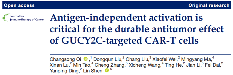
Researchers studied the humanized single-chain variable fragment (scFv) YM01 and YM02 targeting GUCY2C. Among them, YM01 demonstrated stronger binding ability to HCT-116-hGCC-L cells with low GUCY2C expression and did not bind to GUCY2C-negative HCT116 cells (Fig. 1a-c). Further construction of CAR-T cells based on YM01 and YM02 showed that YM01 CAR-T cells had higher affinity for recombinant human GUCY2C protein, faster and stronger cytotoxic activity against HCT-116-hGCC-L cells, more cytokine secretion, higher tumor cell killing efficiency after multiple rounds of chronic stimulation, greater cell proliferation, and were more effective in eradicating tumors in vivo with longer persistence (Fig. 1d-j), indicating that YM01 may have an advantage in recognizing tumor cells with low GUCY2C expression.
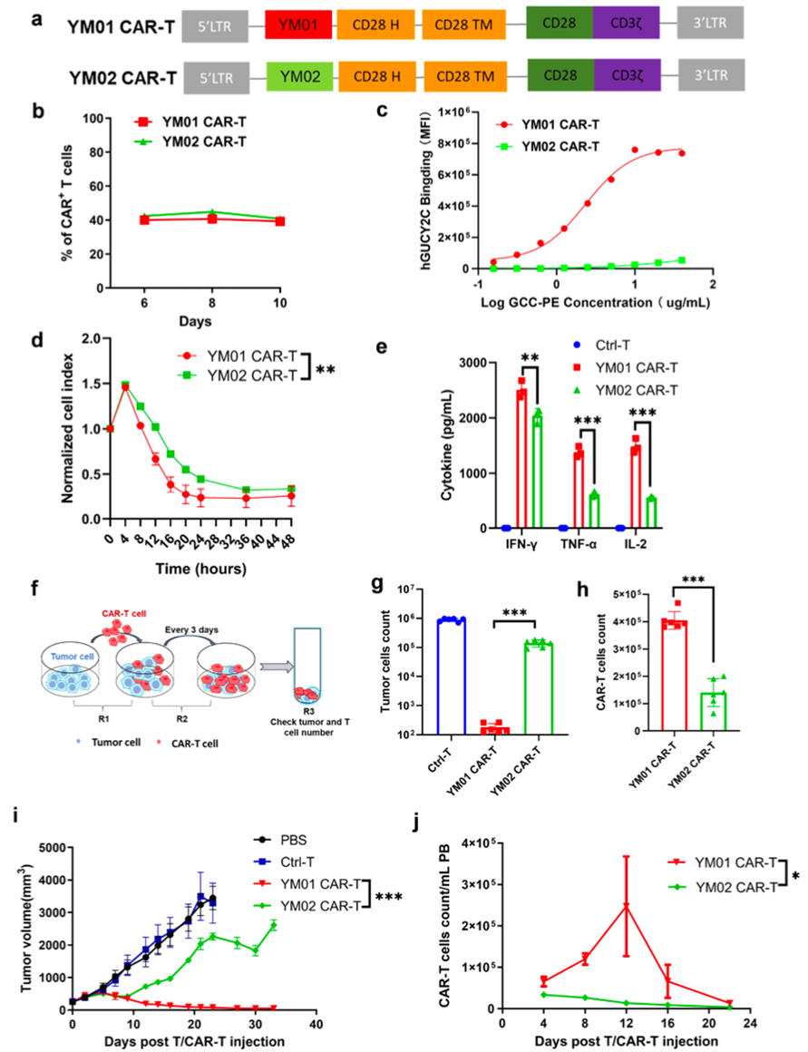
Figure 1. YM01 CAR-T cells exhibit enhanced affinity for GUCY2C and specific cytotoxicity at low effector-to-target cell ratios (E:T).
(a) Schematic diagram of the retroviral vectors encoding YM01 CAR-T and YM02 CAR-T. (b) Representative flow cytometry analysis of the transduction efficiency of YM01 CAR and YM02 CAR in primary T cells at different time points. (c) Binding curves of YM01 CAR-T and YM02 CAR-T cells with hGUCY2CECD-hFc, detecting CAR affinity for GUCY2C by flow cytometry staining with purified PE-labeled hGUCY2CECD-hFc. (d) Real-time cytotoxicity assay of YM01 CAR-T and YM02 CAR-T cells against HCT-116-hGCC-L using RTCA in vitro at an E:T ratio of 1:1. (e) IFN-γ, TNF-α, and IL-2 levels in the supernatant were measured separately using a Cytometric Bead Array (CBA) kit after 24 hours of co-culture. (f) Protocol for repeated chronic stimulation assays. Tumor cells were seeded in 48-well plates one day before T cell addition. On day 0, CAR-T cells were added at a T cell to tumor cell ratio of 1:3. Every 3 days, all T cells were collected and transferred to new wells that had been seeded with 1×10⁵ HCT-116-hGCC-L cells one day prior. (g) Residual HCT-116-hGCC-L cells were collected and counted by flow cytometry during the third round of co-culture. (h) CAR-T cells were collected and counted by flow cytometry during the third round of co-culture. (i) Tumor size was measured over time. (j) The number of CAR-T cells (CD3⁺CAR⁺) in peripheral blood of mice from each treatment group was detected over time by flow cytometry.
03 The Impact of the CD28 Co-stimulatory Domain on the Anti-tumor Function of YM01-based CAR-T Cells
The costimulatory domain is an important component of the chimeric antigen receptor (CAR) structure, which is a domain capable of providing additional signals for T-cell activation. Researchers compared the functional differences of CAR-T cells when using CD28 and 4-1BB as costimulatory domains, and found that CAR-T cells with the CD28 costimulatory domain exhibited superior performance in certain aspects (such as anti-tumor activity against cells with low GUCY2C expression). The researchers constructed 28z and BBz CAR-T cells containing YM01scFv, CD8α hinge and transmembrane domains, and CD3ζ intracellular domain, with either CD28 or 4-1BB costimulatory domains respectively, and both CAR expressions increased over time. Under various experimental conditions, 28z CAR-T cells containing the CD28 costimulatory domain outperformed BBz CAR-T cells with the 4-1BB costimulatory domain in terms of cytotoxicity against tumor cells with low GUCY2C expression, proliferation, cytokine secretion, and persistence.
04 The Impact of Hinge Domain Length on the Effector Function of YM01-CD28-CAR-T Cells and the Role of the CD8α Transmembrane Domain
The length of the hinge domain refers to the number of amino acids contained in the hinge region of the CAR molecule. The hinge domain connects the antigen-binding domain and the transmembrane domain, playing a crucial role in the functional performance of CAR-T cells. Researchers constructed four CAR-T cell variants containing different hinges (CD8α, CD28, IgG4-derived) and transmembrane domains (sourced from CD8α or CD28). The IgG4HCD28TM cells showed impaired effector function, while the other three groups demonstrated high tumor clearance activity. Among them, the CD8HCD8TM group had the fewest residual tumor cells and the highest number of surviving CAR-T cells (Fig. 2a-f). Modifying the IgG4 hinge domain can restore related cellular functions, and a longer hinge domain is critical for cellular effector function (Fig. 2g-j).
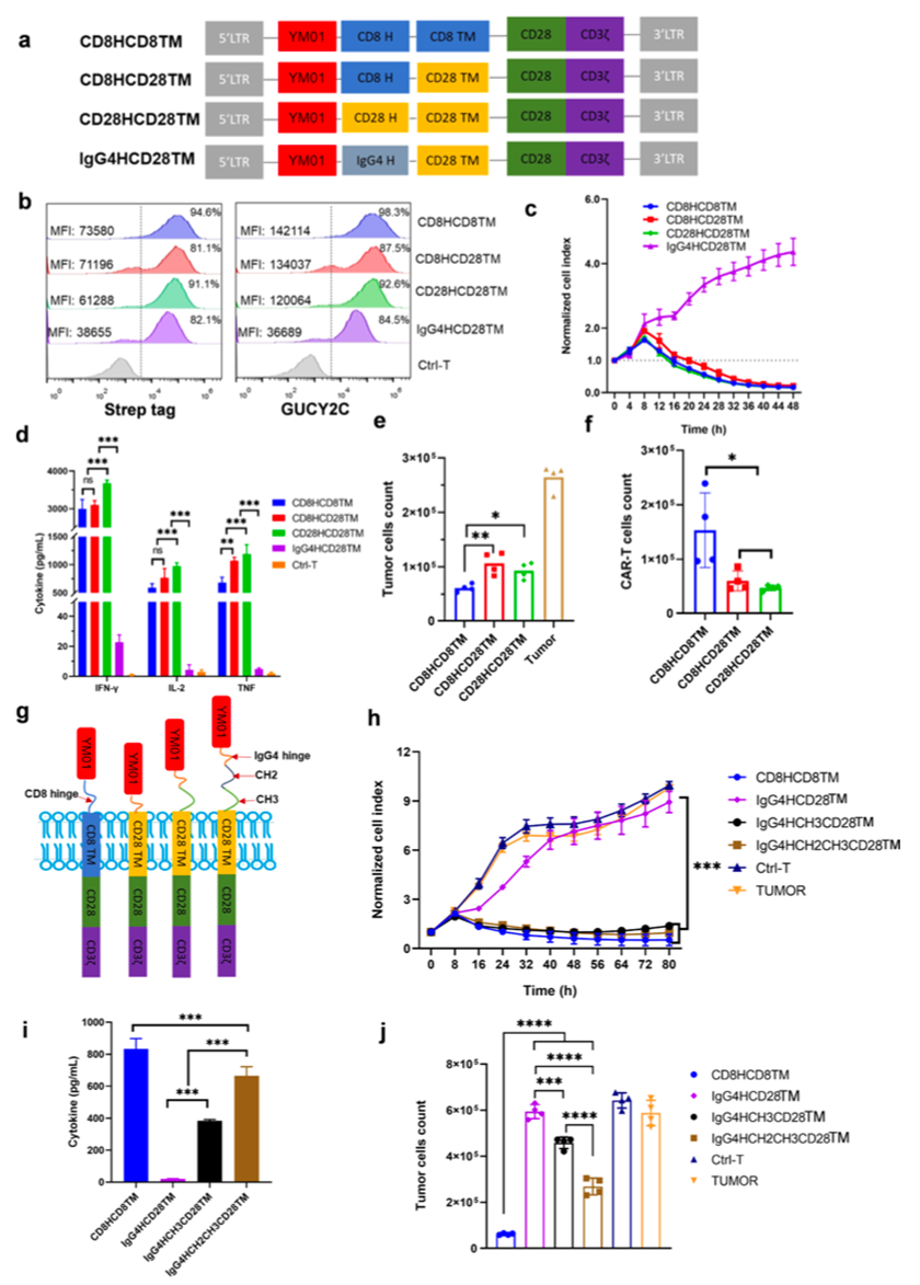
Figure 2. YM01 CAR-T containing the CD8 hinge region and CD8 transmembrane domain demonstrates excellent antitumor efficacy in vitro.
(a) Schematic diagram of the retroviral vector encoding different H and TM domains of the YM01 CAR construct. (b) CAR expression in four YM01 CAR-T cells and their binding to GUCY2C were measured on day 12 by StrepTagII expression or PE-labeled GUCY2C antigen, respectively. (c) Real-time cytotoxicity assay of four GUCY2C CAR-T cells against HCT-116-hGCC-L was detected in vitro at an E:T ratio of 1:3 using RTCA. (d) CAR-T cells were co-cultured with HCT-116-hGCC-L at an E:T ratio of 1:3, and after 24 hours, IFN-γ, TNF-α, and IL-2 in the supernatant were measured using a CBA kit. (e) Residual HCT-116-hGCC-L cells were collected and counted at the 4th round of co-culture. (f) The number of surviving CAR-T cells was collected and counted at the 4th round of co-culture.(g) Schematic diagram of YM01-IgG4 hinge (H) based CARs with spacers of different lengths. All YM01-IgG4 hinge-based CARs use the CD28 transmembrane domain. (h) Real-time cytotoxicity assay of four GUCY2C CAR-T cells against HCT-116-hGCC-L in vitro at an E:T ratio of 1:3 using RTCA. (i) Measurement of IFN-γ secretion in the supernatant after 24 hours of co-culture with HCT-116-hGCC-L cells. (j) Residual HCT-116-hGCC-L cells were collected and counted during the third round of co-culture.
05 The Impact of CD8α-Derived Hinge and Transmembrane Domains on Moderate Signaling in YM01-CD28-CAR-T Cells
Tonic Signaling Evaluation of YM01-CD28-Based CAR-T Cells with Different Hinge and Transmembrane Domain Combinations in the Absence of Antigen and Dynabeads StimulationIn the absence of antigen and Dynabeads stimulation, researchers evaluated tonic signaling for YM01-CD28-based CAR-T cells constructed with different combinations of hinge and transmembrane domains. It was found that IgG4HCD28TM had the lowest activation marker levels when CAR-T cells were activated without antigen, indicating minimal self-activation without external stimuli. In contrast, CD8HCD8TM cells exhibited the highest activation levels, with the highest proportions of effector memory T cells and effector T cell subsets. These cells could survive and proliferate even without interleukin-2 (IL-2) and demonstrated the strongest proliferation and cytokine secretion capabilities. After extending the hinge domain of IgG4HCD28TM CAR-T cells, cells such as IgG4HCH2CH3CD28TM were generated. Experimental results showed that IgG4HCH2CH3CD28TM cells significantly improved self-activation and proliferation abilities, suggesting that CD8α-derived domains are beneficial for moderate signaling and that the length of the hinge domain is essential (Figure 3). This further demonstrates that characteristics of the hinge domain, including its length, significantly impact tonic signaling and related functions of CAR-T cells. Appropriate modifications to the hinge domain can regulate the self-activation and proliferation capabilities of CAR-T cells.
Figure 3. Tension signaling of YM01CAR-T cells with different hinge (H) and transmembrane domains (TM) during in vitro expansion.
(a) Expression of activation and exhaustion markers in four types of YM01CAR-T cells after Dynabeads removal at day 8. (b) Memory T cell phenotype of the four YM01CAR-T cells. (c) Proliferation of human YM01CAR-T cells with different hinge and TM domains during in vitro expansion without cytokine supplementation. (d) T cells were harvested from cultures, washed, and reseeded at 1×10⁶/mL. Supernatants were collected after 24 hours of culture and cytokines were detected using a CBA kit. (e) Activation markers CD25 and 4-1BB in IgG4-based hinge CAR-T cells. (f) In vitro proliferation of IgG4-based hinge CAR-T cells 4 days after Dynabeads removal.
06 The Impact of CD8α Hinge Domain Cysteine Residues on Moderate Signaling and Persistent Function of YM01-CD28-based CAR-T Cells
Mutation of the cysteine residue in the CD8 hinge domain of CD8HCD8TMCAR-T cells showed transduction efficiency similar to that of CD8HCD8TM, but with reduced CAR expression intensity and binding affinity to GUCY2C protein, decreased cytotoxic activity and interferon-γ secretion, increased residual tumor cells after chronic stimulation, reduced CAR-T cell numbers, downregulated antigen-independent activation marker expression, and inability to proliferate without IL-2 (Figure 4).
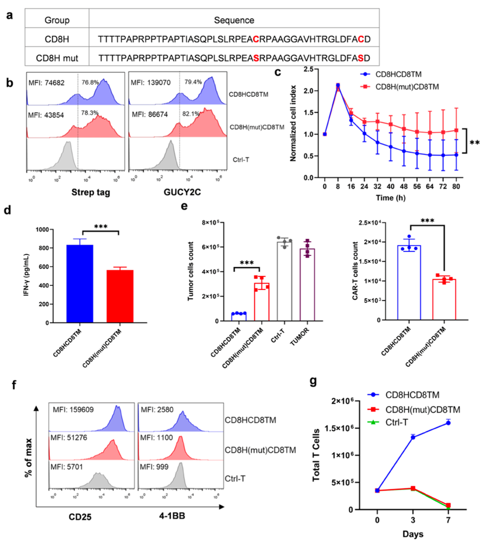
Fig. 4. Tension signaling of YM01-CD8HCD8TM.28z CAR-T cells largely depends on two cysteines in the CD8 hinge.
(a) Mutation of two cysteine residues in the hinge region of CD8HCD8TMCAR. (b) CAR expression on CAR-Ts and binding to GUCY2C measured by StrepTagII expression or PE-labeled hGUCY2C antigen on day 10. (c) Real-time cytotoxicity assay of CAR-T cells against HCT-116-hGCC-L in vitro at an E:T ratio of 1:3 using RTCA. (d) CAR-T cells co-cultured with HCT-116-hGCC-L at an E:T ratio of 1:3, and IFN-γ in the supernatant measured by CBA kit after 24 hours. Residual HCT-116-hGCC-L cells and CAR-T cell counts collected and counted during the second round of co-culture. (f) Expression of activation markers on CAR-T cells 8 days after Dynabeads removal. (g) In vitro proliferation of human CAR-T cells 4 days after Dynabeads removal.
07 YM01-CD28-based CAR-T Cell Anti-tumor Activity in CRC Organoids and In Vivo
● Organoid Experiment:The researchers selected CRC organoids with low GUCY2C expression for the experiment (Figure 5a). Tumor organoids were treated with CAR-T cells or mock T cells (mockT cells, generally used as a control) and over time, the intensity of caspase-3/7 in tumor cells and the infiltration of CAR-T cells into the organoids were evaluated. The results showed that CAR-T cells rapidly surrounded the CRC organoids and effectively infiltrated them after 48 hours. Compared with the mockT group, the fluorescence intensity of caspase-3/7 in CRC organoids was stronger in the CAR-T group after 72-120 hours of co-culture (Figures 5b, c).
● In vivo experiments (patient-derived xenograft models):The study established a patient-derived xenograft (PDX) model of colorectal cancer (CRC) in immunodeficient NCG mice using CRC tissues expressing GUCY2C (Figure 5d). Tumor-bearing mice were injected with 5 million mock T cells or CAR-T cells. The results showed that tumors in mice injected with CAR-T cells rapidly regressed, and the anti-tumor effect persisted during the follow-up period (Figure 5e).
In summary, YM01-CD28-based CAR-T cells with CD8α-derived hinge and transmembrane domains effectively eradicated primary CRC tumors both in vitro in CRC organoid models and in vivo in patient-derived xenograft models that more closely resemble real clinical conditions, even when GUCY2C was expressed at low levels. This further demonstrates the potential of these cells in cancer therapy.
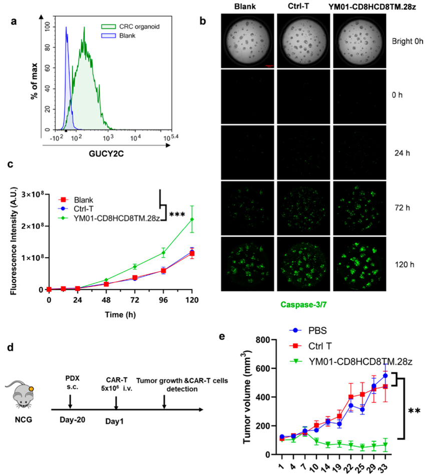
Figure 5. YM01-CD8HCD8TM.28z CAR-T cells exhibit potent effector functions in vitro and in vivo.
(a) Flow cytometry analysis of GUCY2C expression in colorectal cancer organoids. (b) Organoids were collected and counted prior to CAR-T treatment assessment via brightfield and immunofluorescence imaging. Representative single slices of brightfield and caspase-3/7 (green) images from co-culture of YM01-CD8HCD8TM.28zCAR-T with organoids at an E:T ratio of 1:3. (c) Normalized caspase-3/7 signals collected at different time points post co-culture for each treatment group. (d) Timeline of in vivo tumor experiments using patient-derived xenograft (PDX) tumor models. Five million CAR-T cells were intravenously injected into mice 20 days after tumor implantation. (e) Tumor size measured over time.
03
Research Inspiration
1. Organoids More Accurately Simulate the Tumor Microenvironment
Organoids are derived from tumor tissues and can retain some biological characteristics and tissue structures of tumor cells in an in vitro three-dimensional culture system. Compared with traditional two-dimensional cell culture models, they can more accurately simulate the microenvironment of tumors in vivo. This allows researchers to study the interaction between CAR-T cells and tumor cells under conditions closer to the physiological state, providing a more clinically relevant experimental model for evaluating the anti-tumor effects of CAR-T cells.
2. Screening for Highly Efficient Anti-Tumor CAR-T Cell Candidates
By co-culturing CAR-T cells with organoids, and observing the killing effect of CAR-T cells on tumor cells within the organoids as well as the infiltration ability of CAR-T cells into the organoids, the functionality of CAR-T cells can be assessed more intuitively. In this study, this method helps to screen CAR-T cells with high anti-tumor activity, providing more promising candidate cells for further clinical applications.
3. Make personalized medicine possible
Organoids can be established from tumor tissue samples of patients, providing a potential avenue for personalized medicine. The tumor cells of different patients exhibit heterogeneity in gene expression, antigen expression, etc. Using organoid models, the efficacy of different CAR-T cells can be tested based on the tumor-specific characteristics of each patient, thereby offering personalized treatment options.

Scan the QR code to download the original text.
IBAC®(Integrated Biomimetic Array Chip) O2 is a dynamic co-culture organoid chip. It achieves pump-free, high-throughput dynamic fluid perfusion driven by a precision shaker gravity system; the co-culture of organoids with stromal cells or organoids from different tissue sources enhances the biomimicry of the model, creating a more realistic platform for drug screening and toxicology testing. (For more details, please click the link below.)
Organoid Chip Series (IV): Dynamic Co-culture Organoid Chip IBAC® O2
Author: Summer | Reviewer: Rachel | Designer: Rebecca