"Vitreal Tornado" in Sickle Cell Retinopathy Assessed by Widefield OCTA: A Case Report from International Users of Tuopai Medical's 400k-Speed Swept-Source OCT System
TowardPi
High-end Ophthalmic Medical Device Developer


As TowardPi steadily enters the international market and gains increasing favor from international ophthalmology experts, clinical research and article publications have followed in quick succession. Recently, Dr. Marco Rispoli and Dr. Marta Gilardi from the Rome Ophthalmological Hospital in Italy used TowardPi's 400,000-speed ultra-widefield full-region swept-source OCTA Bei Ming · Kun to observe a rare case of sickle cell retinopathy (SCR). The case report, titled "“Vitreal Tornado” in Sickle Cell Retinopathy Assessment by Widefield Optical Coherence Tomography Angiography (Ultra-Widefield OCTA Evaluation of 'Vitreal Tornado' in Patients with Sickle Cell Retinopathy)," was published inAmerican Journal of Ophthalmology》Journal.
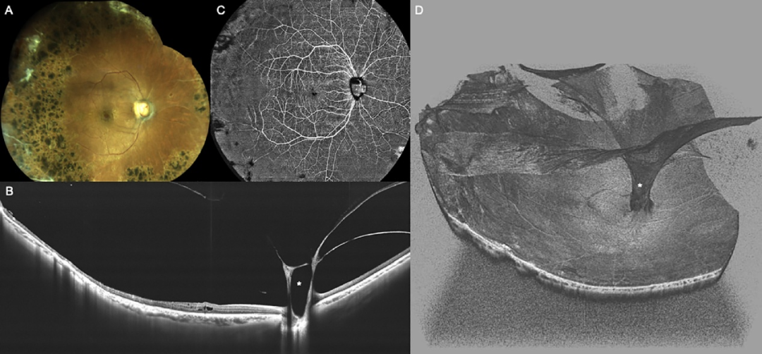
A 61-year-old African American female patient was diagnosed with SCR. She underwent argon laser retinal photocoagulation 20 years ago. Color fundus photography revealed old laser spots on the peripheral retina of her right eye (Fig. A, color fundus montage). Ultra-widefield swept-source OCT examination detected vitreous abnormalities: grade 3 posterior vitreous detachment in the right eye, with part of the dense vitreous tightly connected to the optic disc (indicated by the asterisk in Fig. B). A 24mm x 20mm ultra-widefield OCT angiography (OCTA) image of the superficial retinal vascular network clearly showed typical ischemic changes in the peripheral retina associated with SCR, accompanied by capillary dropout and large areas of non-perfusion (Fig. C). Through ultra-widefield 3D OCT reconstruction of the posterior pole, the cortical vitreous connected to the optic disc was observed to extend upward in a funnel shape, resembling a "tornado" sweeping through the fundus (Fig. D), vividly reproducing the extremely rare and unique vitreous characteristics of this patient.
SCR is an ocular complication of Sickle-cell disease (SCD). Its pathological process involves the aggregation of abnormal hemoglobin in red blood cells within the retinal microcirculation, leading to reduced deformability of red blood cells. As the condition progresses, retinal vascular changes occur, including peripheral arterial occlusion, peripheral arteriovenous anastomosis, secondary neovascularization, secondary vitreous hemorrhage, and severe complications such as tractional or rhegmatogenous retinal detachment.400,000-speed ultra-widefield full-range swept-source OCTA captures 120° high-definition ultra-widefield imaging in a single shot within 7-15 seconds. Combined with rich vascular quantification parameters, it easily evaluates vascular lesions and peripheral retinal changes caused by diseases and can reconstruct ultra-widefield fundus tissues three-dimensionally.,better assist clinical in-depth qualitative and quantitative research on diseases!


Article Writing:Li Xin, TowardPi Medical
Content Proofreading:Jian Zhou, TowardPi Medical
TowardPi Reading Series:
Answer Revealed! Can Ultra-Widefield OCTA Replace Fluorescein Angiography for Diabetic Retinopathy?
Not Only a Substitute, but a Superior Choice! Wide-Angle Swept-Source OCTA Replaces FFA for RVO!
New Evidence Added! Ultra-Widefield OCTA Can Replace Traditional Angiography in Diabetic Retinopathy
Massive Subretinal Hemorrhagic PED Secondary to PCV: Rapid Postoperative Resolution, AJO Report
Advancements in Swept-Source OCT and Swept-Source OCTA (English Translation)
"Dolphin" Leaping Out of the Retina — A Case of Congenital Vitreous Cyst
A Stunning Visual Journey: Ultra-Widefield OCTA Unveils Choroidal Changes in Thyroid Eye Disease
OCTA Reveals Sensitive Association Between Retinal Arteriole Diameter and Mean Arterial Pressure
— Scan the QR code to follow us —


WeChat Official Account
WeChat Video Channel

Every like and view you give, I sincerely take as a sign of affection.