Boyun Xundao: Swept-Source Intraoperative OCT-Guided Microscope Enhances Precision and Safety in Glaucoma Surgery
TowardPi
High-end Ophthalmic Medical Device Developer
Glaucoma is the world's leading irreversible blinding eye disease, and surgery is one of its important intervention methods. Preferred procedures such as glaucoma drainage device implantation and minimally invasive glaucoma surgery (MIGS) often face limitations like restricted field of view and insufficient visibility, usually relying on the subjective experience of the surgeon. Additionally, critical surgical steps, such as the insertion depth and positioning of the drainage device in the anterior chamber (whether it touches the corneal endothelium or iris), as well as the creation of space under the scleral flap, can directly impact surgical outcomes if there are intraoperative deviations.
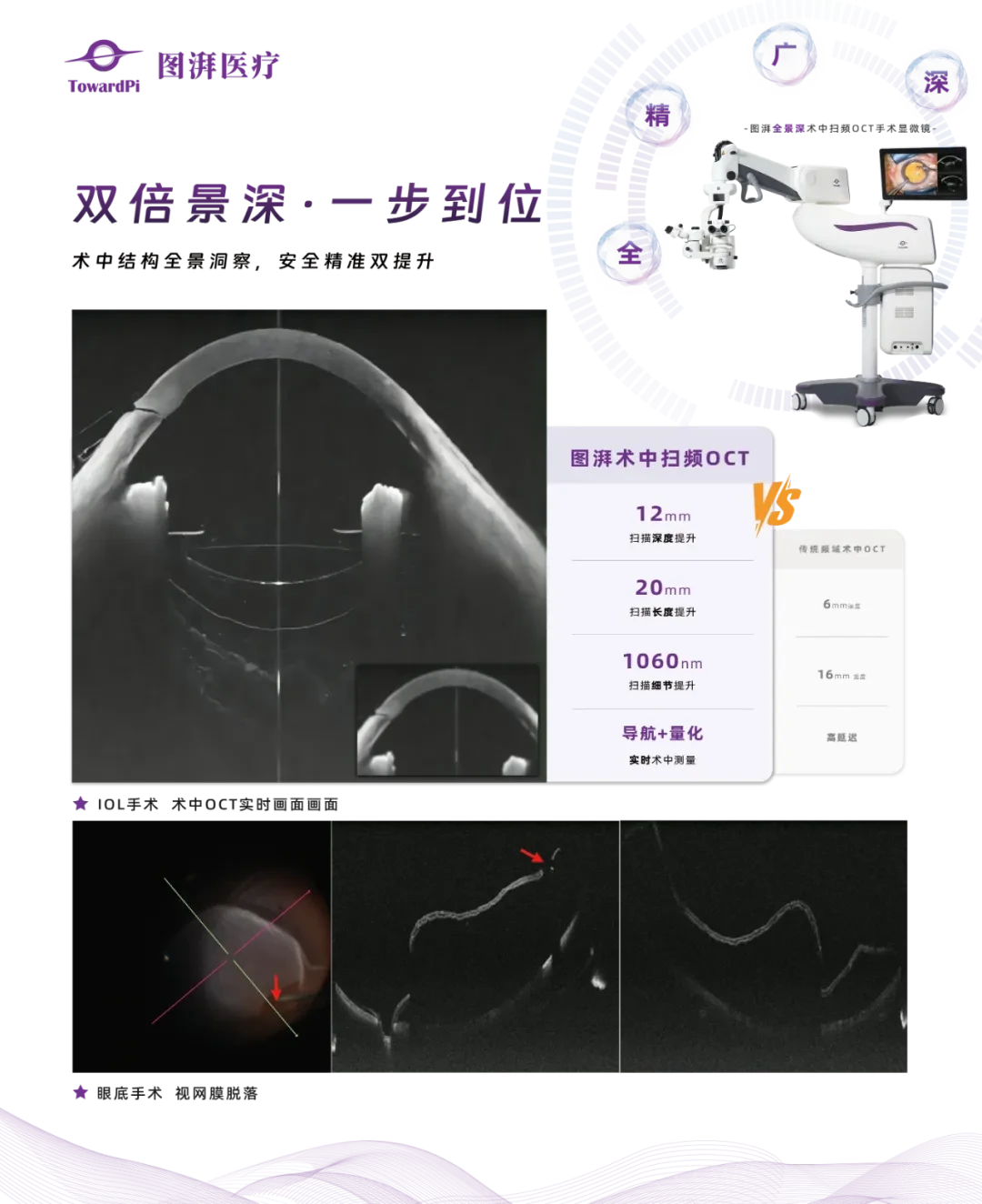
TowardPiSwept-Source OCTOCT(SS-iOCT)Navigational MicroscopeFor SurgeonsProvided real-time high-definition intraoperativeOCT Image,With a "transparent", precise, and quantifiable third perspective, the medical risks caused by "blind operations" are eliminated. Professor Zhang Han and his team from Shandong First Medical University Affiliated Provincial Hospital were the first to utilize real-time SS-iOCT imaging for seamless observation, guidance, and evaluation of glaucoma surgery in a clinical setting.
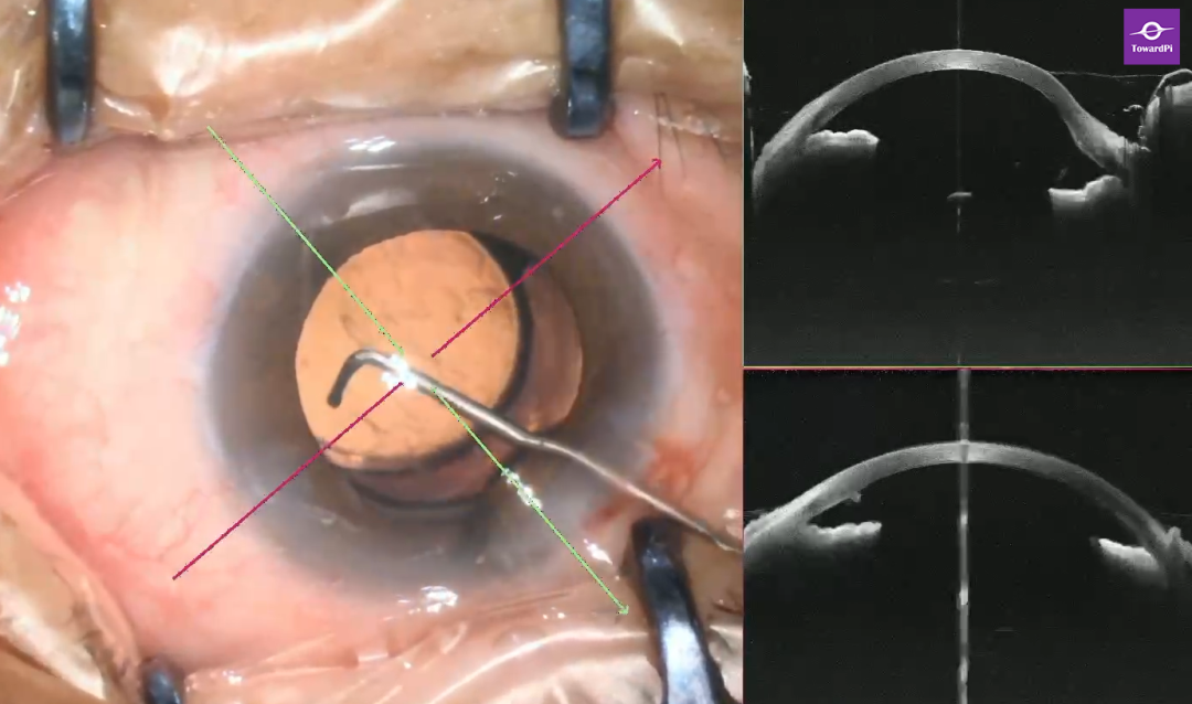
Case 01: SS-iOCT-Guided Angle Drainage Device Implantation
Diagnosis: Optic Atrophy (Right) Pseudophakia (Right)
Preoperative Examination: Intraocular Pressure39mmHg Visual Acuity 0.2 (Decimal Visual Acuity Chart, hereinafter the same)
Imaging Report:
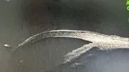
(a) Intraoperative SS-iOCT-guided puncture positioning
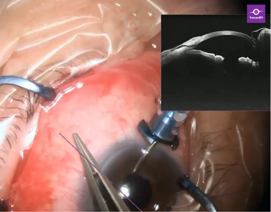
(b) After puncture, use SS-iOCT to quantitatively analyze the length of the three-segment drainage device and observe the drainage status.
Postoperative Review: IOP 11mmHg Visual Acuity 0.2
DiseaseExample02: SS-iOCT-Guided Goniosynechialysis
Diagnosis: Chronic Angle-Closure Glaucoma (Right)
PreoperativeExamination: Intraocular Pressure38mmHg Visual Acuity 0.7
ImagingReport:
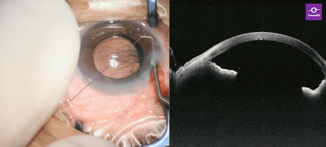
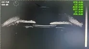
(b)Before Angle Separation
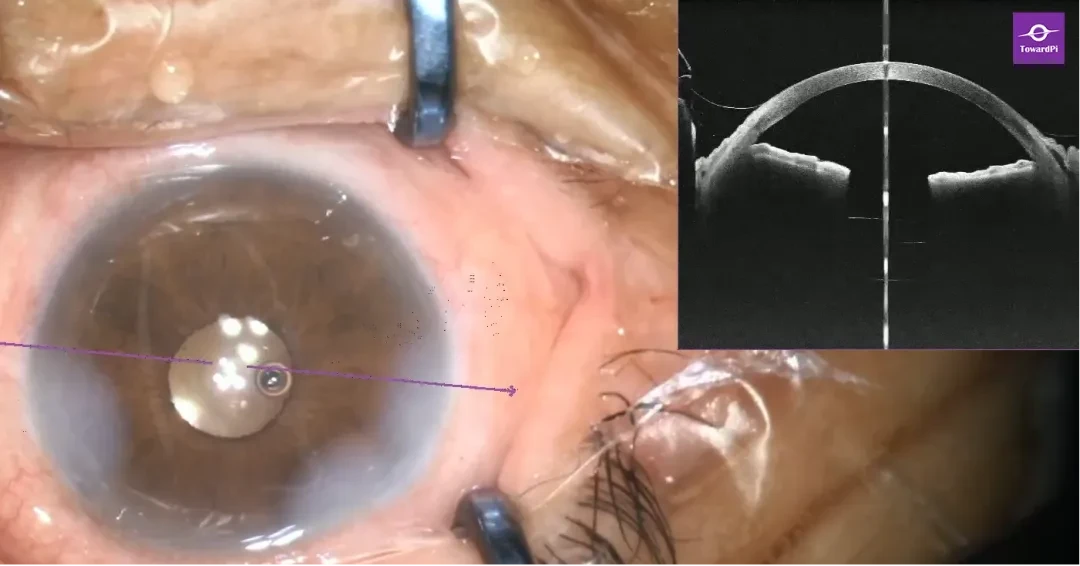
(c)After goniosynechialysis
Postoperative Review: Intraocular Pressure16mmHg Visual Acuity 0.7
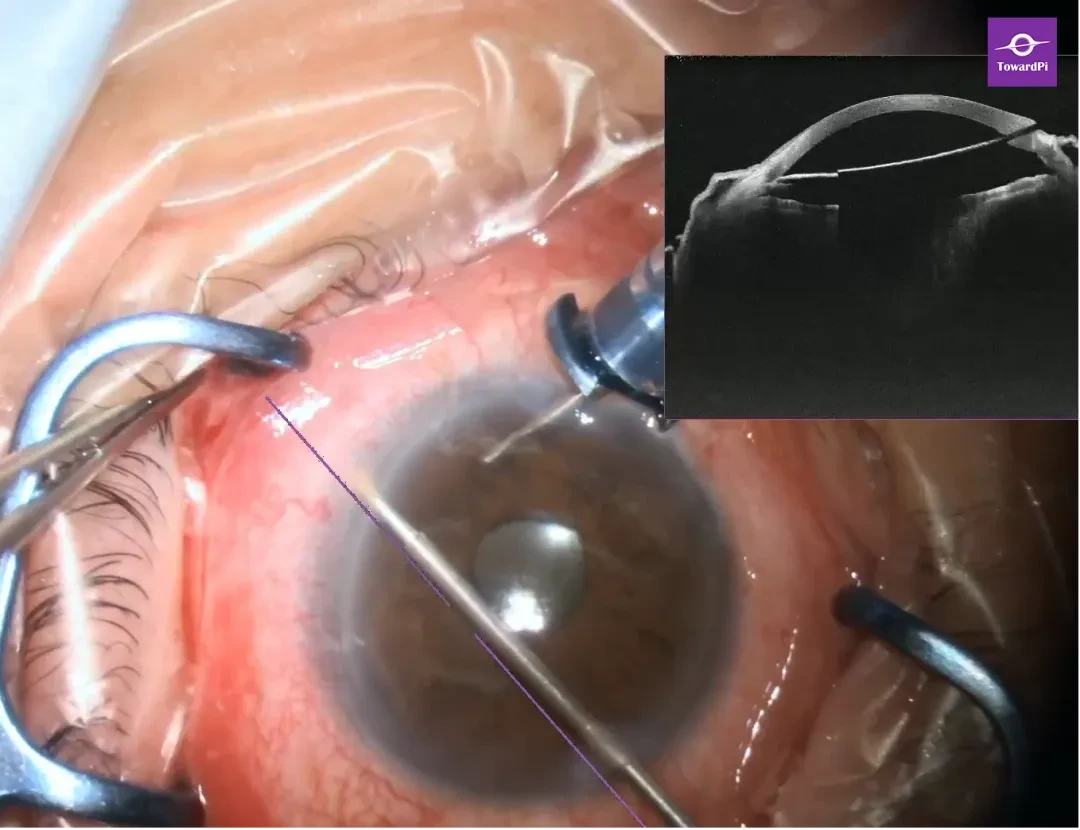
Professor Zhang Han believes that the swept-source intraoperative OCT navigation microscope can during the surgical procedure:
Provide cross-sectional images in real time.
High-resolution display of microscopic structures such as the cornea, limbus, iris, and drainage devices.
Assist doctors in performing the most precise operations.
With the advancement of technology and the increasing clinical demands, innovative surgical techniques and products for glaucoma are also rapidly evolving. The multi-modal fusion imaging navigation of TowardPi's surgical microscope and SS-iOCT supports more precise, safer, and successful implementation of innovative surgical techniques and drainage device implantations, helping to achieve better patient outcomes.
Technological Innovation, Together Guarding the Light!
Expert Introduction


Zhang Han
Director of Ophthalmology, Provincial Hospital Affiliated to Shandong First Medical University, Medical Doctor, Chief Physician, Doctoral Supervisor
Specializing in the diagnosis and treatment of various types of complex cataracts, and functional refractive reconstruction for ocular trauma.
Served as a member of the Ophthalmology Branch of the Chinese Medical Association
Member of the Cataract Group of the Chinese Medical Association
Member of the Ophthalmology Branch of the Chinese Medical Doctor Association
Deputy Secretary-General of the Ophthalmology Professional Committee of the Chinese Medical Doctor Association's Postgraduate Education
Chairman of the Ophthalmology Branch of Shandong Provincial Medical Association


Pan Hong
Deputy Chief Physician of Ophthalmology, Shandong First Medical University Affiliated Provincial Hospital, Medical Doctor
Committee Member of the Fundus Disease Group, Chinese Society of Microcirculation
Deputy Group Leader of the Youth Group, Ophthalmology Branch, Shandong Medical Association
Member of the Cataract Group, Ophthalmology Branch, Shandong Medical Association
Vice Chairman of the Ophthalmology Branch of Shandong Young Medical Workers Association
Vice Chairman of the Ophthalmology Innovation and Development Branch of Shandong Research Hospital Association
Standing Committee Member of the Ophthalmology and Visual Science Branch of Shandong Research Hospital Association
Member of the Ocular Trauma Professional Group of the Shandong Provincial Association of Rehabilitation Medicine Vision Rehabilitation
Proficient in the diagnosis and treatment of refractive errors, cataracts, optic nerve disorders, vitreoretinal diseases, and endophthalmitis, with particular expertise in surgical treatments for cataracts, vitreoretinal diseases, and ocular trauma.
Led and participated in 2 National Natural Science Foundation projects, led and participated in 4 provincial-level projects in Shandong Province, and published more than 10 SCI papers and Chinese Journal series papers.