Intalight's Ultra-Widefield OCTA Featured on Cover of Journal Français d’Ophtalmologie, Marking Fourth International Journal Cover Recognition

Intalight
High-end Ophthalmic Equipment Developer
Intalight Literature Issue 162——Recently,FromIntalightIntalight Full-Eye如意OCT
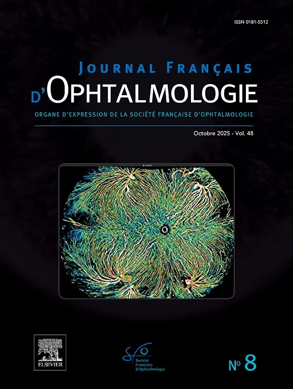
Another Scientific Research AchievementReturn to FranceOphthalmology Journal Cover,225°Ultra-Wide AngleOCTAShowcasing Intalight's Globally Leading Imaging Capabilities。Journal Français d’Ophtalmologie《French Journal of Ophthalmology》2025Year10Monthly Journal (Vol.48 No.8) The cover image is provided byFrench Fundus Imaging ExpertAdil EL MaftouhiTeamProvided, showcasing the Ruyi Full-EyeOCTUltra-Wide AngleOCTAInChoroidal Blood Flow ImagingInnovative Breakthroughs and Outstanding Performance. This is the fourth time that Intalight's full-eye OCT imaging has been featured on the cover of an international journal, marking the continued international recognition of Intalight's technical strength and academic influence.
Intalight Past Journal Covers:
Intalight's OCTA Imaging Featured on the Cover of Ophthalmology! Case Report on Retinal Capillary Hemangioma
French Fundus Imaging Expert's Case Featured on the Cover! Intalight OCTA Displays Neovascularization in Fine Detail
Published on the cover of the Eye journal! Case sharing of retinal arteriovenous malformation
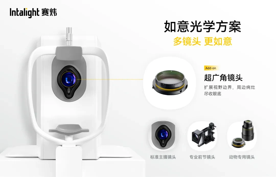
Intalight Full-Eye OCT: Pioneering Multi-Lens Combination DesignMacular and Optic Disc Fine Imaging, Ultra-Wide Angle Fundus Imaging, Panoramic Anterior Segment Imaging and Animal ImagingFour different scenarios with dedicated lenses to avoid the performance compromise of "one lens for all," making every examination more satisfactory.Among them,Ultra-Wide Angle LensEven more, through a significant expansion of the field of view, achievingComprehensive Observation of Extensive Lesions, LetPeripheral Lesions Clearly Visible。
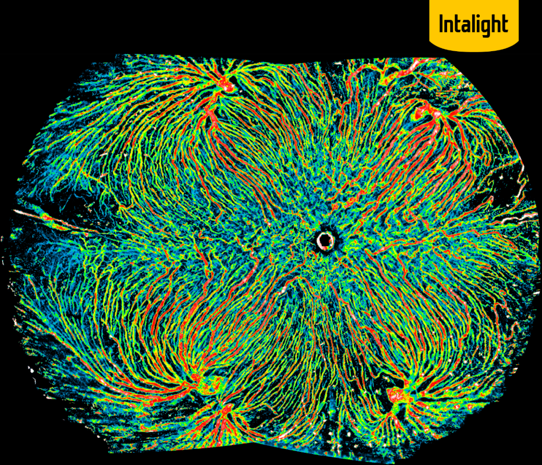
The cover image of this issue is provided byIntalight Full-Eye OCTAcquisition, Based onSingle 26 mm×21 mm Ultra-Widefield OCTA Scan, throughFive-direction Automatic Mosaic,Achieved up to225° Ultra-Widefield Choroidal Blood Flow ImagingIn this image, multiple choriocapillaris ampullary-like structures are clearly visible in all four quadrants, with blood vessels fully exposed. This provides an unprecedented panoramic view for the clinical evaluation and scientific research of choroidal diseases.

Adil EL Maftouhi
With over 20 years of experience in ophthalmology, he is currently an expert in ophthalmic imaging at the Paris 15-20 Hospital in France, and also works at the Rive Ophthalmology Center in Geneva, Switzerland, as well as the Paris Eye Research Institute. His expertise and clinical research focus on anterior and posterior segment imaging technologies, with particular proficiency in OCT and OCTA techniques. He has authored numerous peer-reviewed publications and book chapters on these topics, and is the author or co-author of several books on ophthalmic imaging. He has delivered speeches at numerous national and international conferences and is a co-organizer of the Lyon Retina Academic Conference.
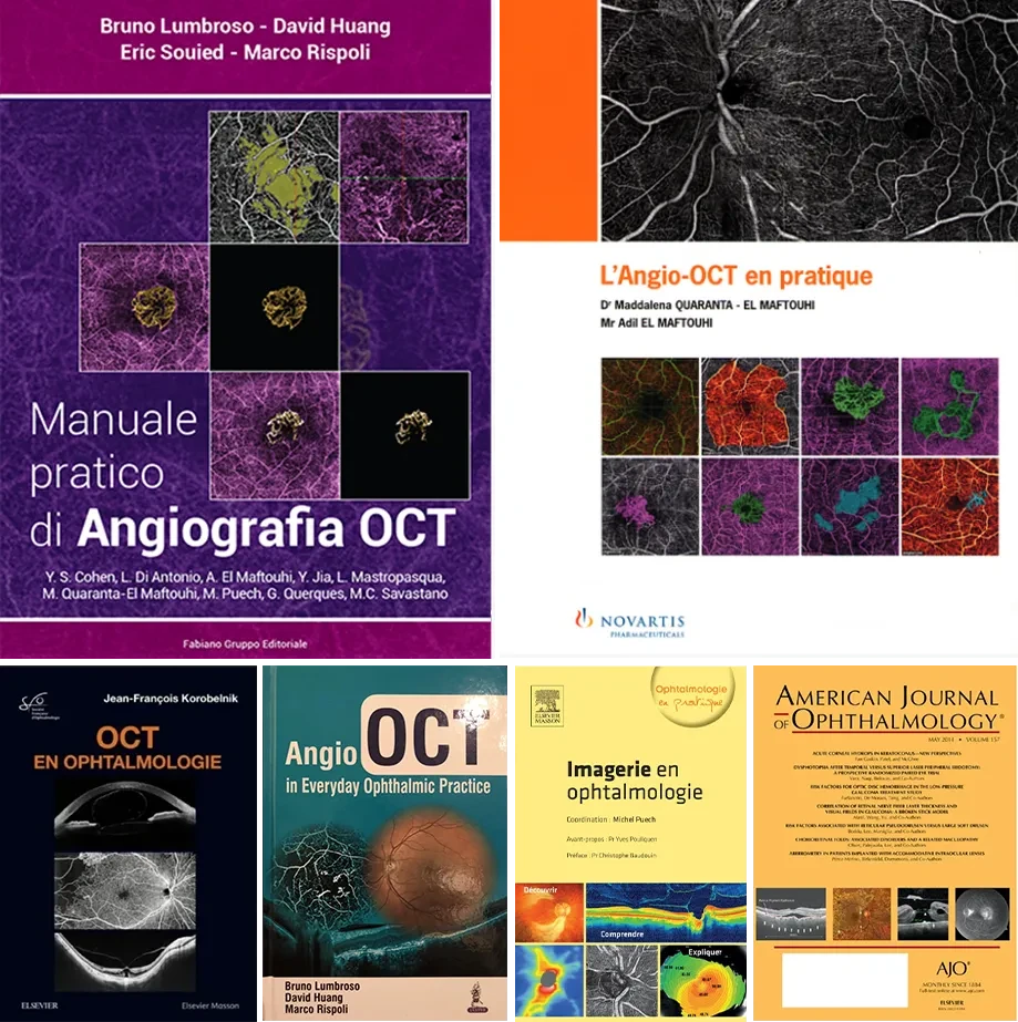
Books on Ophthalmic Imaging Partially Compiled by Adil