TuPai Medical's Wang Yingqi: Pioneering Intraoperative 3D OCT Navigation and Exporting High-End Ophthalmic Equipment to Europe
TowardPi
High-end Ophthalmic Medical Device Developer


Ophthalmic Surgery: Navigating the "Blind Dive" with Precision
The eye, with a diameter of less than 3 centimeters, is the most intricate optical system in nature.
In the past, if doctors wanted to "repair" this system, they could only use a regular "magnifying glass" to perform surgeries within a narrow space using a two-dimensional perspective. The exact nature of the lesions beneath the ocular surface could only be inferred based on experience.
"TowardPi: Breaking New Ground in Medical Innovation"Wang Yingqi, a "post-90s" young entrepreneur from Tsinghua University, collaborated with the university’s research team to co-found TowardPi. Their development of swept-source OCT (Optical Coherence Tomography) ophthalmic devices has broken international monopolies. Moreover, the world's first intelligent surgical microscope featuring real-time 3D OCT navigation during operations is at the forefront globally. These devices seem to bestow doctors with "X-ray vision," revealing deep ocular lesions with unprecedented clarity and significantly enhancing the precision of intraoperative decision-making.
Now, high-end ophthalmic diagnostic and therapeutic equipment with Beijing's innovative genes is rapidly making its way to the global market.
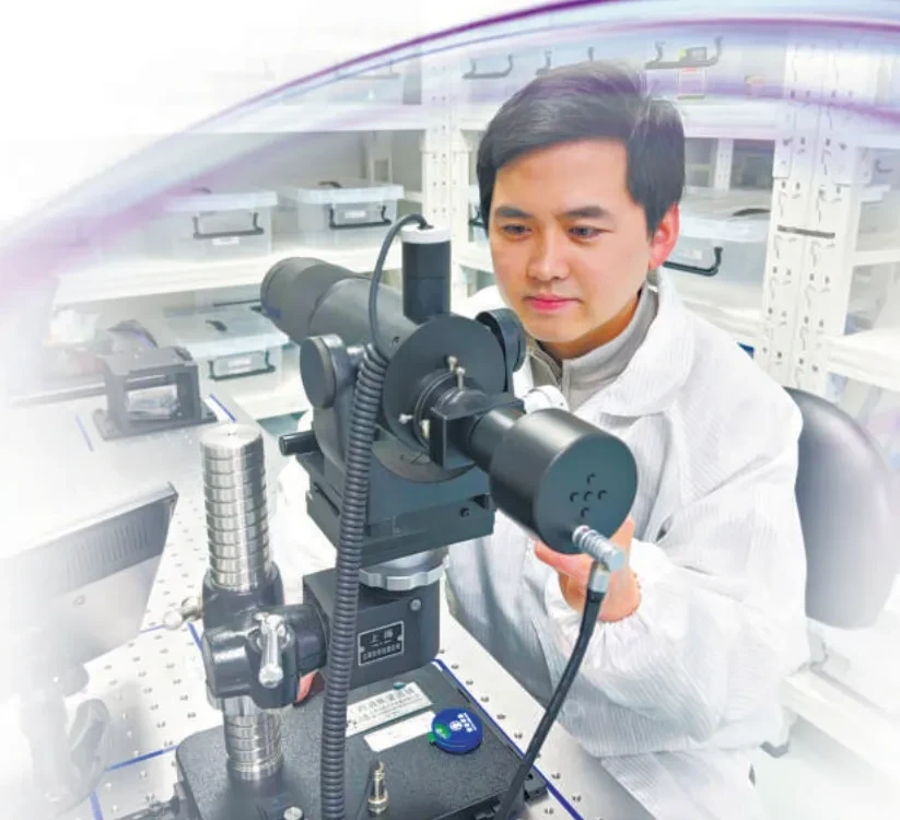
Wang Yingqi Working in the Lab
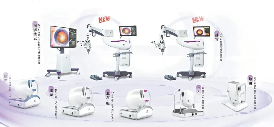
TowardPi Full Series Product Images
· "Blind Diving" in a Tiny Space
The macular region of the human eye, which is responsible for central vision and fine visual details, has a diameter of only a few millimeters and a thickness of less than 0.5 millimeters, yet it contains approximately 6 million cone cells. These cells convert light signals into neural electrical signals that are transmitted back to the brain. Once the macular region is damaged, only ophthalmologists with skills as delicate as "embroidery" are capable of preserving the patient's remaining vision.
In general surgical operations, an accuracy level in millimeters is sufficient to meet the requirements of most procedures. However, in the macular region of the retina, the precision of surgical instruments must reach a level of 10 micrometers, equivalent to one-seventh the diameter of a human hair. This extreme demand for precision stems from the fragility and intricacy of the macula, which is the core area responsible for central vision and fine visual details. Any minor iatrogenic trauma can lead to severe damage to visual function. Nevertheless, surgeons capable of performing such "microscopic-level" surgeries are extremely rare. In China, there are over 45,000 ophthalmologists, but less than one-tenth of them are qualified to perform complex vitreoretinal surgeries, indicating a significant talent gap. One of the key reasons for this shortage is the limited field of view provided by surgical tools.
"Traditional ophthalmic surgical microscopes are essentially a special kind of 'magnifying glass' that can only help doctors see enlarged flat images. As for the deep ocular structures beneath the plane, they rely on the doctor's clinical experience to analyze and judge," said Wang Yingqi, CEO of TowardPi. This experience-dependent operating model places extremely high demands on the professional competence of doctors.
"Beneath the surface, hidden currents surge."
An abnormal bulge in the fundus, a region with unusual color—whether it’s edema, hematoma, cyst, or tumor—doctors typically rely on a combination of preliminary imaging results, subtle tactile feedback from surgical instruments during the operation, as well as the appearance of the affected area, the patient's age, medical history, and other factors. Clinical experience is key to navigating the "hidden obstacles" during surgery and completing the delicate task of "defusing" the lesion. Wang Yingqi recalled the scene while observing a surgery: "Sometimes, when doctors face a lesion of unknown nature, in order to accurately determine its type, they may need to spend more than half an hour on repeated attempts. A complex surgery often takes three to four hours."
In addition to facing重重困境in decision-making, the精细复杂的解剖结构of the eye also greatly tests the surgeon's操作手感. Ocular lesions are often紧密交织with surrounding blood vessels and nerves. Relying on two-dimensional images to construct a three-dimensional anatomical structure in the mind and then precisely performing surgery is often too difficult for young doctors. The lack of intuitive 3D imaging technology also means that surgical outcomes cannot be assessed promptly and must be judged through follow-up observations several days post-surgery. If some surgical outcomes do not meet expectations, a second surgery may be required, which is a heavy burden for both doctors and patients.
Intraoperative navigation equipment is the key solution to this problem.
Current common CT technology (X-ray computed tomography) can present three-dimensional images of specified areas inside a patient's body through large-area scanning. However, in the field of ophthalmology, CT technology is difficult to apply — CT imaging has only sub-millimeter resolution and cannot reconstruct ocular structures with high precision.
In 2017, Wang Yingqi discovered the key to improving resolution. That year, in the laboratory of Professor Huo Li's team from the Department of Electronic Engineering at Tsinghua University, he saw the prototype of swept-source OCT for the first time. "At first glance, it looked like a piece of bone," Wang Yingqi recalled with a laugh. "Although its appearance was crude, I was amazed by its imaging speed, quality, and probing depth. When the prototype was placed close to the skin, even the minute structures beneath could be seen clearly."
Actually, as early as 2014, Wang Yingqi had attempted to promote the transformation of scientific and technological achievements in the field of medical imaging. "During my graduate studies, I mainly focused on bioluminescent molecular imaging research. At that time, I realized that the true value of a scientific research achievement should not just remain in papers, but should be transformed into tangible and valuable results." However, reality was fraught with difficulties. In the following year, under the guidance of his supervisor, Wang Yingqi worked alone using the limited equipment available in the lab for research and development, but it ended in regret. "Technical principles are only one part of the process of transforming achievements; subsequent product development, production implementation, and other stages are all crucial and indispensable."
The collision between biomedical engineering and optoelectronic technology has sparked a whole new "chemical reaction."
In a small 20-square-meter house outside the southeast gate of Tsinghua University, Wang Yingqi and Huo Li, along with their team, began to grapple with that "bone." At that time, the transformation of original scientific achievements in China's medical device field had not yet reached scale, with few cases to reference. However, Wang Yingqi set a clear and resolute goal: complete the development of the engineering prototype within three months and obtain the medical device license within one and a half years.
The confidence stems from a completely new technical route of swept-source OCT. Previously, the Huo Li team had already validated the underlying system principles, and core components such as lasers, high-speed acquisition cards, and photodetectors had preliminarily completed lab-level R&D, making it seem like everything was ready.
The team achieved its first goal as scheduled, with the engineering prototype's test performance reaching an internationally leading level. However, when the prototype was handed over to doctors, the feedback left Wang Yingqi feeling disheartened: "The parameters are indeed impressive, but this equipment would only work if the patient were a wooden figure."
"As a medical device for patients, we completely ignored ergonomic design—a cold and bulky iron frame supporting the head, and any slight movement by the patient requires the doctor to readjust the handle; the software interface is cluttered with hundreds of operation buttons, leaving doctors disoriented and struggling to make precise adjustments." Wang Yingqi admitted that this "setback" taught the team a crucial lesson: "At the time, the engineering team fell into a misconception, believing that solid device performance was enough. They never considered things from the perspective of doctors and patients, neglecting their experience and feelings during use."
· Make diagnosis "final and decisive"
Without understanding the needs of both doctors and patients, they made every effort to learn. Wang Yingqi and the R&D team immersed themselves in the hospital, spending time with doctors during consultations whenever possible, observing how doctors examined and operated on patients, and identifying the difficulties and bottlenecks in the process.
The working principle of OCT is to use light as a "sampling needle" to quickly penetrate tissues and extract images in an extremely short period of time. Therefore, the faster the scanning speed and the greater the depth, the larger the range and the clearer the image that can be obtained within the limited shooting time. In the eyes of doctors, ophthalmic detection devices based on OCT technology available on the market at that time were not sufficient in terms of scanning speed and depth, and could not provide a definitive diagnostic conclusion, but could only be used for auxiliary examinations.
Taking diabetic retinopathy as an example, doctors need to obtain images of the entire area from the edge to the center of the retina to complete the diagnosis. However, due to the slow scanning speed and insufficient imaging depth of OCT devices at that time, the single-shot range was too small. Therefore, the most commonly used clinical examination method remains angiography.
"Some patients are allergic to contrast agents and need to be observed for about an hour, which slows down the department's examination rhythm significantly—only a few patients can be examined in a day." Curious, Wang Yingqi asked the doctor why not use OCT technology to complete the examination through image stitching? The doctor immediately invited a patient to cooperate for a demonstration. Since these patients were generally older and suffered from visual field loss, they were unable to follow the doctor’s instructions to move or fixate their eyes. "Throughout the entire morning, we tried with more than ten patients, and only one patient’s captured images could barely be stitched together—but the result was still fragmented and unsuitable as a basis for diagnosis or treatment."
Wang Yingqi realized that in the development of medical devices, underlying technologies must be combined with clinical needs to achieve transformation. At the same time, he also discovered that even mature OCT products in the international market have parameters such as scanning speed and range that still do not meet the "gold standard" requirements for ophthalmic diagnosis. As a result, he completely overturned the initial prototype design concept and continuously raised the R&D goals for his team: the bandwidth rate of the photoelectric detector conversion needed to be increased, the scanning laser had to be further accelerated, and the data acquisition card had to process massive amounts of data at high speed...
Faster, even faster. Behind the goal lies the need for fundamental technological innovation.
Among the components, the data acquisition card, which is only the size of a palm, was the most challenging part in the development of the entire system. "It serves as both the hub for data aggregation and the 'brain' of the entire device, involving thousands of components, four layers of circuit boards, and a massive amount of embedded code," said Wang Yingqi. From the outset, Wang Yingqi and the team were determined to pursue self-developed innovation, collaborating with the technology transfer team from the Institute of Microelectronics of the Chinese Academy of Sciences to develop a domestically produced board. Throughout this process, they faced challenges at every turn, from software and hardware design to heat dissipation structures, and from signal filtering to electromagnetic compatibility.
Many interfaces of domestically produced boards are not yet mature. During testing in a hospital environment, the device may encounter error reports ranging from 10 to 20 times a day, indicating that compatibility still requires ongoing optimization. The significant increase in computing power has also led to heat dissipation challenges, causing the system to "shut down" when overheating. Moreover, the faster the computing speed and the greater the bandwidth, the higher the noise level—when electronic products such as mobile phones or tablets get slightly close, the screen immediately displays a snow-like image.
This research and development effort, which started from scratch, was persisted in for 3 years by Wang Yingqi and the team. "At the most frustrating times, I also struggled with whether to buy imported finished components. But even the best imported boards could only achieve one-third of our required speed. We couldn't rely on anyone else, so we had no choice but to grit our teeth and keep going." Fortunately, the feedback from doctors using the product improved day by day, giving the team the motivation to continue.
Finally, in 2019, Beijing welcomed the first innovative medical device application in the field of ophthalmology. Wang Yingqi's team completed 200 clinical trials in just three weeks at Peking Union Medical College Hospital and Tsinghua Changgung Hospital. Both the image quality and various parameters of the equipment received high praise from the review experts.
"Compared with similar products in the international market, our improvements are not just 'minor tweaks' but a comprehensive leap in all parameters," Wang Yingqi proudly introduced the outstanding performance of this swept-source OCT device: it can complete 400,000 scans per second, which is more than four times faster than imported flagship products at that time; the maximum detection depth has increased from the traditional two to three millimeters to 14 millimeters, capturing deeper tissue details more clearly; the imaging angle has expanded from 50 degrees to 120 degrees, covering a wider field of view... The operation is as simple as a 'one-touch' system, allowing patients to complete the examination in just 10 seconds with their eyes open, significantly improving diagnostic efficiency.
The comprehensive and significant enhancement of equipment functionality also facilitated the successful inclusion of ultra-high-speed, ultra-wide-angle blood flow imaging technology in China's ophthalmic diagnostic guidelines the following year, establishing itself as the "gold standard" for clinical diagnosis. Currently, this equipment has achieved 100% coverage across the top 300 ophthalmic hospitals in China, with the total domestic installed base surpassing 1,000 units, ranking first in market share within the industry.
What makes us even prouder is that such market share was not achieved through a low-price strategy. Relying on leading performance, TowardPi’s products are priced higher than similar imported equipment, yet they still gain widespread recognition from clinicians." Wang Yingqi’s tone was full of pride as he explained that this recognition is inseparable from the collaborative innovation between the team and frontline doctors during the R&D process. "In real clinical scenarios, our equipment significantly improves diagnostic efficiency for hospitals and optimizes the patient experience. This is the core value of the integration of medicine and engineering."
· Install "navigation" for surgical knives
The product was an instant hit, but Wang Yingqi's team did not stop there. They set their sights on a route that no one worldwide had yet achieved: integrating ultra-high-speed swept-source OCT technology into ophthalmic microscopes to provide real-time "navigation" for ophthalmologists during surgery. They also aimed to increase the resolution of ophthalmic surgical microscopes from sub-millimeter to micrometer levels, making surgical operations more precise and safer.
"The intraoperative swept-source OCT navigation microscope is suitable for almost all ophthalmic surgeries." Wang Yingqi used a straightforward analogy to highlight the core value of this technology: "It’s like driving a car. In the past, you had to drive a manual transmission, and beginners were always distracted by shifting gears and pressing the clutch, often losing focus on the road. But our device is like an automatic car, freeing doctors from tedious operational concerns so they can concentrate fully on the surgical details and execute each step with precision."
To achieve this route, the difficulty is also unprecedented. As an inspection device, OCT allows for a certain amount of computational delay; even if it takes one or two minutes to process the data, it will not affect the inspection results. However, when used for surgical navigation, the requirements are extremely strict — once the delay exceeds 100 milliseconds, the image will have motion blur, directly affecting the safety of the surgery, which is an impassable "red line." Using swept-source OCT technology to complete the acquisition, storage, computation, and rendering of large-scale 3D images within tens of milliseconds is an extremely challenging goal that has never been achieved before.
The R&D journey was fraught with challenges. The feasibility study of the project lasted nearly half a year. After the team initiated the R&D, problems came pouring in: the performance of the data acquisition card needed further enhancement, the volume of data processing increased significantly, the optomechanical structure became more complex, and the number of components in the overall device was five times that of previous swept-source OCT equipment. "More challenging was the interaction and interference between components during integration—almost every component had to be redesigned and repeatedly tested," recalled Wang Yingqi. At the time, some industry insiders frankly stated that the R&D goals for the equipment were set too high, appearing somewhat "overambitious."
"Every project we initiate, every challenge we tackle, is about pushing the limits and turning the impossible into the possible." Wang Yingqi admitted, the team always adheres to one belief: either don't do it, or do it to the extreme. "Fortunately, we've won every battle!" In 2025, the world's first intelligent surgical microscope with real-time 3D swept-source OCT navigation functionality received Class II medical device certification from the National Medical Products Administration (NMPA) in China, officially launching into the market.
Wang Yingqi will never forget the moment when the first OCT navigation microscope was installed. "Several engineers and I pushed the equipment into the hospital lobby, personally unpacked the outer packaging, cut the seal, and completed the installation step by step," he recalled. "When the first device entered the hospital, many doctors were still skeptical and reluctant to let it into their operating rooms. However, by the time the second device was about to be installed, the operating rooms were vying to extend invitations, hoping it would be prioritized for installation in their rooms."
Nowadays, in cataract surgery, the patient's corneal endothelium, anterior and posterior capsules, zonules status, and anterior chamber can be stably presented in real-time, significantly improving the precision of operations such as capsulorhexis and nucleus division. In vitreoretinal surgery, abnormal conditions like edema, cysts, and tumors are clearly distinguished, assisting doctors in precisely controlling the depth and range of resection. In surgeries for retinal vascular diseases, real-time blood flow information is displayed during the operation, providing reliable support for surgical decision-making...
With the help of atlas-based, quantitative three-dimensional images, doctors no longer need to spend effort "guessing" at lesions. This not only helps young doctors grow faster but also ensures that the chief surgeon can focus entirely on the operation itself—every procedure is purposeful.
In January 2026, the National Healthcare Security Administration updated the charging guidelines, and intraoperative swept-source OCT navigation was included as an independently chargeable item, benefiting an increasing number of patients. "Ophthalmic surgeries cannot be delayed; one day earlier in surgery means one day earlier in halting the progression of the disease, giving patients more hope for recovery," said Wang Yingqi. The advancement of OCT technology has enabled more ophthalmologists to perform complex surgeries, which is the most practical benefit for grassroots patients.
In more cutting-edge fields, Wang Yingqi's team is advancing with new plans. This time, their target is a neurosurgical operating microscope project, aimed at meeting the real-time navigation needs of brain-computer interface surgeries. "For brain-computer interface systems to achieve more complex functions, it is necessary to obtain sufficient and high-precision EEG signals, which requires the precise implantation of high-throughput electrodes. Intraoperative OCT navigation will play a crucial role in this," said Wang Yingqi. Compared to ophthalmic surgeries, neurosurgical operations require lasers with longer wavelengths and equipment with higher power. "All key components need to be upgraded, and many technical difficulties cannot be resolved by referring to previous literature; only persistent exploration can pave the way."
After nine years of entrepreneurship, TowardPi Medical has gone through "three relocations" and ultimately chosen to establish its roots in the Changping International Medical Device City. "The research and development of high-end medical equipment relies heavily on upstream resources. Changping is one of the regions with the highest number of medical device companies in Beijing and also benefits from strong university resources, which greatly assist in R&D and production. Currently, we have basically achieved mass production of our devices," said Wang Yingqi. TowardPi's ophthalmic OCT devices not only cover major medical institutions within China but are also sold in bulk overseas. By 2025, international sales are expected to exceed 10 million US dollars, with 70% being exported to developed European countries such as Germany, Italy, and France.
Not long ago, Beijing's first intelligent ophthalmology medical engineering industrial park — Zhongguan Village (Changping) Intelligent Ophthalmology Medical Engineering Industrial Park — was also established at the Changping International Medical Device City. TowardPi, as one of the co-initiating units, participated in the platform construction, project introduction, and project incubation of the park. "I hope that in the future, this place will become a true innovation base, not just focusing on one or two products, but promoting more international innovative achievements and creating clinical value," said Wang Yingqi.
Source: Beijing Daily, Beijing Association for Science and Technology
Further Reading
1
2
3
4
5