Professor Wan Jia's Team at Yunnan University Affiliated Hospital: Balloon Angioplasty for Thromboangiitis Obliterans of the Lower Extremity Using DKutting™ Scoring Balloon

DK Medtech
Vascular Interventional Balloon Product Developer

With the vigorous development of China's peripheral vascular intervention market, common balloons tend to cause complications such as excessive vascular injury, flow-limiting dissections, and hematomas when treating the increasing number of highly resistant stenotic lesions. In contrast, pressure-focusing balloons utilize cutting/notching elements positioned between the vessel wall and the balloon’s outer diameter during expansion, enhancing localized pressure for efficient directional dilation. This reduces vascular elastic recoil and represents a new direction in the evolution of vascular intervention balloons.
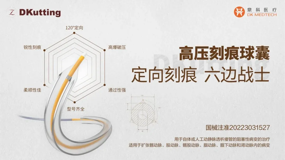
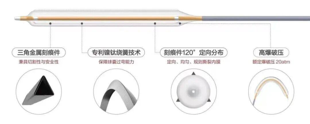
DK Medtech's independently developed DKutting™ High-Pressure Scoring Balloon, featuring an exclusive patented design (CN201810478242.X), boasts numerous advantages such as excellent trackability, uniform expansion, and high burst pressure. In terms of overall product performance, it is referred to as the "hexagonal warrior," marking a significant breakthrough for local enterprises in innovating and surpassing top-tier imported products.
DK Medtech Special Release[Professor Wan Jia's Team: Balloon Expansion for Lower Limb Thromboangiitis Obliterans] Case Presentation, demonstrating the meticulous operation of each case and the clinical application of advanced equipment and instruments. From the formulation of treatment strategies for different cases, standardized intraoperative procedures and technical applications, complication prevention, to perioperative management, etc., the aim is to promote the standardization of diagnosis and treatment for vascular stenosis and occlusive diseases, strengthen technical exchanges and experience sharing among doctors, with the hope of providing new ideas and methods for future diagnosis and treatment, benefiting more clinical patients.

Balloon Expansion for Lower Limb Thromboangiitis Obliterans

Yunnan University Affiliated Hospital, Wan Jia
Patient Information
Basic Information:Male, 71 years old.
Chief Complaint:Coldness and numbness in the right lower limb for over a month.
History of Present Illness:The patient reported that one month ago, without obvious cause, he experienced coldness and numbness in the right lower limb, which worsened after activity and slightly relieved after rest. There was no swelling or pain in the right lower limb, and thus it did not attract attention nor receive treatment. Later, the coldness and numbness in the right lower limb gradually worsened, accompanied by skin pigmentation and anterior tibial ulcer. Today, for further treatment, the patient came to our outpatient department. The lower limb vascular ultrasound suggested "incomplete thrombosis of the right popliteal artery could not be ruled out." The outpatient department admitted the patient with "thromboangiitis obliterans of the right lower limb." Since the onset of the disease, the patient’s mental state, diet, and sleep have been fine, with normal bowel movements, and there has been no significant change in weight.
Past Medical History:Denies history of coronary heart disease, diabetes, and hypertension.
Physical Examination:Pigmentation and ulceration in the right lower limb boot area, with the ulcer crusted over, approximately 1.5cm x 1.5cm in size. The skin temperature of the right lower limb boot area is low, with cyanosis of the toes, and the pulse of the right posterior tibial artery cannot be palpated.
Admission Diagnosis:
Thromboangiitis Obliterans of the Right Lower Limb
Varicose Veins in the Left Lower Limb
Previous interventional treatment
Time | Main Treatment Process |
January 22, 2024 | Lower extremity vascular ultrasound performed during the outpatient visit showed thickened wall, narrowed lumen, and solid echo in the distal part of the right popliteal artery (incomplete thrombosis of the right popliteal artery could not be excluded, further examination recommended), and the patient was admitted to the hospital. |
January 22, 2024 | Lower extremity CTA indicates occlusion of the right popliteal artery and arteriovenous fistula in the right lower limb. |
January 23, 2024 | Treated with clopidogrel, beraprost sodium tablets, and atorvastatin calcium tablets. |
January 24, 2024 | Right femoral artery puncture under local anesthesia, right lower limb arterial angiography + right popliteal artery balloon angioplasty + right lower limb arteriovenous fistula coil embolization. |
January 30, 2024 | Improved and discharged from the hospital, continued oral treatment with clopidogrel, beraprost sodium tablets, and atorvastatin calcium tablets after discharge. |
March 4, 2024 | The patient has ulcerated and crusted lesions on the right lower limb, without pain or coolness discomfort in the right lower limb. |
Preoperative Analysis
Preoperative Analysis:
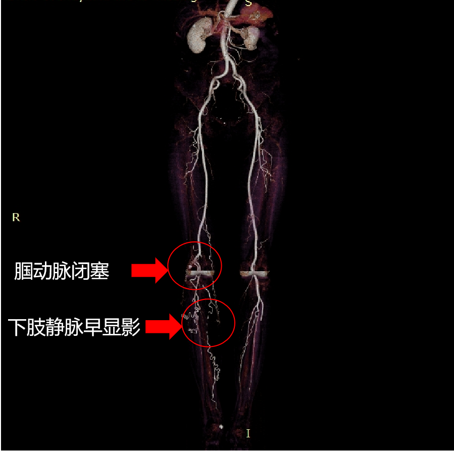
Preoperative CTA showed:Occlusion of the right popliteal artery with collateral circulation formed around it; early visualization of the veins in the right lower leg, suggesting the formation of an arteriovenous fistula.
The patient has occlusive disease in the right popliteal artery, with a high risk of dissection after plain balloon dilation. The lesion is located at the joint, and stent placement after dilation may have long-term patency that remains to be verified. Consider using a scoring balloon to directionally relieve the pressure of the hyperplastic intima and reduce the occurrence of flow-limiting dissections. Combined with a drug-coated balloon, this approach ultimately aims to minimize the need for graft placement.
Surgical Objective:
Main Objectives:Open the right popliteal artery and embolize the right lower limb arteriovenous fistula as much as possible.
Secondary Objective:Open the right popliteal artery and place a stent at the same time.
Surgical Strategy/Plan:
The right femoral artery was punctured antegradely, and a 6F short sheath was inserted.
A 150cm guidewire was used in conjunction with a single-bend angiographic catheter to pass through the distal segment of the occluded right popliteal artery and reach the anterior tibial artery.
Application of DKutting™ 4.0mmx80mm to dilate popliteal artery occlusive lesions;
Applied drug-coated balloon 5.0mmx150mm to dilate the lesion segment;
Embolization of arteriovenous fistula in the calf segment with micro-coils; 6. Occluder closure of the puncture site.
Surgical Procedure

Preoperative angiography showed occlusion of the right popliteal artery with collateral circulation formation.

The blood supply to the lower right leg is adequate, with early venous imaging.
4.0mmx80mm DKutting™ Scoring Balloon Catheter used for right popliteal artery balloon dilation, with a noticeable notch observed.

Embolization of arteriovenous fistula in the lower right leg.

After pre-dilation, drug-coated balloon angioplasty was performed, restoring blood flow in the popliteal artery with significant improvement in imaging.
Follow-up
Discharge Status:At discharge, the crust of the ulcer on the anterior tibia of the patient's right lower limb had fallen off, the wound was healing well, and there was no discomfort such as pain or coldness in the right lower limb. After discharge, the patient continued oral treatment with clopidogrel, beraprost sodium tablets, and atorvastatin calcium tablets.
Follow-up after 3 months:No pain, numbness, or cold discomfort in the right lower limb; the ulcer has healed well, and the right lower limb is mobile.
Case Summary
Case Characteristics:Elderly patient, chronic course, right lower limb ischemia leading to pain, coolness, and non-healing ulcers in the right lower limb, with occlusive arterial disease in the right lower limb.
Preoperative Assessment Key Points:Preoperative lower limb CTA examination to clarify the location, nature, length of the lesion, and collateral circulation.
Surgical Strategy/Technical Key Points:Select an appropriate balloon and prepare the vessel with a suitable balloon diameter. After completion, use a drug-coated balloon dilation to suppress intimal hyperplasia and extend patency time.
Features/Usage Tips of the Device:The pressure focusing and directional expansion of the scored balloon provide better lumen results, while the drug-coated balloon suppresses intimal hyperplasia.

Expert Introduction


Professor Wanjia
The Surgeon of This Case
Ph.D., Associate Chief Physician, Master's Graduate Supervisor,Director of Vascular Surgery, Affiliated Hospital of Yunnan University;Visiting Scholar at the Division of Vascular Surgery, Stanford University Medical Center, USA;Member of the Society for Vascular Surgery (SVS);Member of the Youth Committee of the International Union of Angiology (IUA);Committee Member of the Thoracic Aortic Disease Committee, China Branch of the International Union of Angiology (IUA);Member of the Vascular Regeneration Group, Tissue Repair and Regeneration Branch, Chinese Medical Association;Vice Chairman of the Vascular Surgery Branch of Yunnan Medical Association;Group Leader of the Abdominal Aortic Disease Group, Vascular Surgery Branch, Yunnan Medical Association;Standing Committee Member of the Vascular Surgery Branch of Yunnan Province Medical Association;Member of the Endovascular Medicine Professional Committee of the Yunnan Province Medical Association;Member of the Interventional Physicians Branch of the Yunnan Province Medical Association.

Department Introduction

The Third Department of General Surgery (Vascular Surgery) of Yunnan University Affiliated Hospital was officially established in January 2005. In 2007, it was officially designated by the Yunnan Provincial Health Department as the Yunnan Vascular Surgery Center. In 2015, the department was approved by the Yunnan Provincial Health Department for the key clinical specialty construction project in Yunnan Province. Since its establishment, after 18 years of development, the department has become a clinical department integrating medical treatment, teaching, and research. It has obtained the standardized training base for resident physicians in Yunnan Province, the standardized training base for specialized physicians in Yunnan Province, the peripheral vascular interventional diagnosis and treatment technology training base in Yunnan Province, and the continuing medical education base in Yunnan Province. It is currently the largest provincial peripheral vascular disease diagnosis and treatment center in Yunnan Province.
The department's scope of diagnosis and treatment includes open surgical treatment and minimally invasive interventional treatment for complex aortic dissection, thoracoabdominal aortic aneurysms, severe lower extremity arterial sclerosis occlusion, diabetic foot, visceral artery occlusion, as well as open and interventional treatment for carotid artery stenosis. It also provides one-stop treatment for deep vein thrombosis of the lower extremities and pulmonary embolism, thermal ablation technology for treating varicose veins of the lower extremities, autologous stem cell transplantation for chronic critical limb ischemia, and comprehensive treatment for congenital hemangiomas and arteriovenous malformations. A specialized nursing clinic has been established, with technical features including wound repair, lymphedema treatment, and PICC catheterization, currently placing it among the advanced technical ranks in the province. The vascular surgery department has gradually developed to form multiple subspecialty treatment teams covering aortic diseases, lower extremity arterial diseases, venous diseases, lymphatic diseases, and wound repair.
The department currently has 35 authorized beds and a total of 24 medical staff, including 9 doctors and 15 nurses. Among them, 4 have doctoral degrees, 4 have master's degrees, and 6 are master's degree supervisors. There are 2 chief physicians, 3 deputy chief physicians, and 1 deputy chief nurse. Two of the department’s physicians are reserve talents in medical disciplines in Yunnan Province. The department leads and participates in multiple projects funded by the National Natural Science Foundation of China and the basic research program of the Yunnan Provincial Department of Science and Technology. Director Jia Wan, Head Nurse Yan Bao, and several key physicians hold positions such as chairpersons and standing committee members in the Vascular Surgery Branch of the Chinese Medical Doctor Association, the Tissue Repair and Regeneration Branch of the Chinese Medical Association, the Vascular Surgery Branch of the Yunnan Medical Association, the Vascular Surgery Physician Branch of the Yunnan Medical Doctor Association, and the Endovascular Specialty Committee of the Yunnan Medical Doctor Association.
Copyright Statement: This platform aims to help medical and health professionals better understand the latest developments in relevant disease areas. The information content published on this platform does not imply agreement with its descriptions or viewpoints, but is merely for providing more information. If there are any copyright issues, we kindly request the rights holders to contact us, and we will address them as soon as possible. The information is solely for medical and health professionals to stay informed, and such information cannot replace professional medical guidance in any way, nor should it be regarded as medical advice. If such information is used for purposes other than staying informed, this platform and the author shall not bear any related responsibilities.Contact email for collaboration:vascular@edoctor.work。