Optos Reaches Global Milestone of 30,000 Installed Retinal Imaging Systems

NIDEK
Ophthalmic Surgical Instrument Diagnostic Equipment Developer

June 25, 2026Optos (Optos plc)Announce,itsRetinal Imaging DeviceGlobal cumulative installed base reaches30,000 units。
In February 2024, Optos disclosed that its global installed base had exceeded 25,000 units; by June 2025, it reported an installed base of over 27,000 units. According to the company’s figures, this represents an increase of approximately 5,000 units over a period of about 28 months. (The company has not disclosed the specific breakdown by model, region, or customer type.)
#Product Gradient Analysis
Optos was founded in 1992 and acquired by Nikon Corporation in 2015 for JPY 48.128 billion, subsequently becoming a wholly owned subsidiary of Nikon.Optos’s current core feature remains single-capture 200° ultra-widefield scanning laser imaging.。
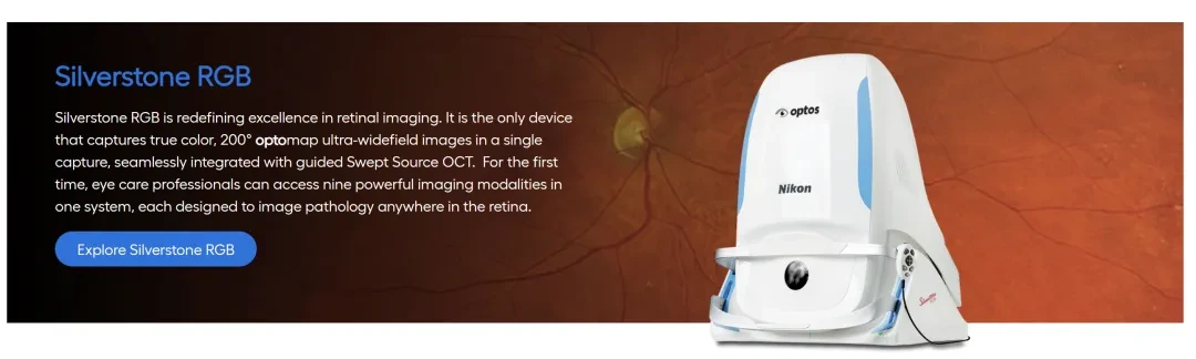
The product portfolio has formed a clear tiered structure.: Daytona is designed for routine fundus examinations, California adds autofluorescence and angiography, MonacoPro integrates spectral-domain OCT, and Silverstone RGB further integrates swept-source OCT, FA, ICGA, and RGB imaging.
The company's 2026 announcement stated that,Its various platforms collectively offer 10 imaging modes.. The latest high-end products will peripheral eyesFundus imaging, OCT, and angiography integrated into a single workstation.
Regarding U.S. regulatory status, the Optos P200TE and P200TxE platforms have received clearance multiple times through the traditional 510(k) pathway. Recent clearances include K231673 and K233602 for the P200TE. The 510(k) determination is based on substantial equivalence to a legally marketed device; it should not be described as PMA approval or broadly referred to as “FDA certification.”
#Comparison of Technical Approaches
Eye Future Search: No Competitors Have Yet Followed the Single 200° Trajectory.
Source: Company official website and FDA database; Search date: June 29, 2026.Manufacturers differ in their definitions of the reference point for the field of view, single-capture imaging, and stitched images. Practical comparisons by hospitals also include peripheral resolution, artifacts, interpretability rate, multimodal configuration, operation time, and after-sales costs.
The advantages of Optos are its single-shot 200° imaging capability and existing installed base.; CLARUS emphasizes true-color RGB and resolution; Mirante highlights the integration of SLO, OCT, and OCTA; SPECTRALIS leverages its existing OCT platform to add ultra-widefield angiography capabilities.
#Clinical Evidence and Usage Boundaries
Optos statesMore than 4,000 device-related papers have been published, with publicly available evidence consistently pointing to two conclusions: ultra-widefield fluorescein angiography can supplement sugarRisk information for diabetic retinopathy; in screening for retinal tears, ultra-widefield images still cannot replace dilated peripheral fundus examination.
Note: Due to differences in study populations, equipment, and endpoints across various studies, direct cross-comparisons are not valid. Relevant studies primarily utilized earlier Optos platforms and therefore cannot automatically demonstrate that the latest models are superior to competing products.This distinction directly impacts product promotion. Research on diabetic retinopathy supports the value of ultra-widefield FA in documenting peripheral leakage, non-perfusion, and lesion distribution.For patients with high myopia, those undergoing preoperative ICL examination, and those with acute posterior vitreous detachment, ultra-widefield imaging demonstrates high specificity but limited sensitivity. While the device can improve the efficiency of documentation, follow-up examinations, and patient communication, it should not replace dilated fundus examination, three-mirror lens examination, or scleral depression.
#Market Competition Landscape
Ultra-widefield scanning laser imaging devices (primarily ultra-widefield confocal scanning laser ophthalmoscopes) market currently presents “Imported Brands Dominate the High-End Market, Domestic Brands Rise Rapidly, and Differentiated Competition Emerges in Niche Segments"landscape."
Imported Brands (Dominating the High-End Market)
Optos(Optos, UK/NIDEK): Utilizes confocal scanning laser ophthalmoscopy (CSLO) technology, providing a 200° ultra-wide field of view in a single capture. NIDEK has launched new models such as Silverstone and Monaco, which integrate OCT functionality.
ZEISS(Zeiss, Germany): Its Clarus series employs true-color wide-field imaging technology, achieving a single-image field of view of 133° and up to 200° through image stitching, maintaining traditional advantages in image quality and high-end multimodal platforms.
Heidelberg(Heidelberg, Germany): Integration into the Spectralis platform via wide-field modules enables combined OCT with wide-field color fundus photography and angiography. In June 2026, the platform received new FDA software clearance for ultra-widefield OCTA imaging.
NIDEK(Nidek, Japan) andTopcon(Topcon, Japan): Holds a certain market share in the field of conventional fundus imaging and some wide-field/ultra-widefield devices, with products renowned for their stability and optical technology. The NIDEK Mirante SLO/OCT is listed as an ultra-widefield platform in industry literature; the Topcon Triton2 was launched in Europe in September 2025.
Domestic Brands (Rapidly Rising, Seizing the Mid-to-High-End and Primary Care Markets)
MicroClear Medical(Suzhou): Its CRO series employs confocal laser scanning technology, with a single-image field of view reaching 150° (this parameter is sourced from corporate promotional materials and has not yet been verified by independent authoritative literature). According to public information, MicroClear Medical launched this series in 2020.
Topcon Medical(Beijing): Leveraging Tsinghua University’s technology, its “Zhuzhao” product employs four-wavelength full-color imaging with a 150° field of view per image, and was launched in April 2024. According to the company, the product features capabilities in multimodal integration and AI-assisted analysis.
Yaoshi Medical(Suzhou): According to the company’s disclosure, it is one of the first enterprises in China to launch a scanning laser ophthalmoscope (SLO) with ultra-widefield confocal laser scanning technology. The Yetsea series utilizes four-laser confocal technology. The Yetsea 300 received National Class II Medical Device Registration approval in July 2024.
Bigwei Medical(Suzhou): The BVF8000 received regulatory approval and was launched in June 2025, featuring a single-image field of view of 170°, a panoramic mosaic field of view of 260°, four-channel multi-wavelength imaging, and an integrated AI-assisted analysis system.
Fuhan Medical(Nanjing): Focused on neonatal/premature infant screening scenarios, 160° ultra-wide-angle pediatric-specific device, lens diameter 7mm, wireless handle weighing 400 grams.
As of the search date, no large-sample, multicenter, peer-reviewed head-to-head studies comparing domestically produced devices with Optos and CLARUS have been identified in publicly available literature. While parameters such as field of view, laser channels, and imaging modes are clearly defined, there is limited disclosure regarding lesion detection sensitivity, readability rates across different quadrants, repeatability, examination time, and actual installed base.
#Eye Future Observation
For domestic companies, the ophthalmic industry looks forward to seeing more validation data in the future, such as:
1. Standardize the field of view and readable area specifications, and disclose imaging quality across different quadrants;
Second, to demonstrate consistency in lesion detection and follow-up through independent image interpretation and multicenter studies;
Third, demonstrate the value of the workflow through examination time, equipment utilization rate, failure rate, and after-sales costs.
For more detailed information, please visit the Optos website:https://www.optos.com/products/

Ophthalmic Solutions Case Studies