Hospitals Leveraging 3D Printing for Complex Medical Interventions: Innovations in Surgical Planning, Custom Implants, and Personalized Care
In the context of manufacturing, compared to other popular industries such as aerospace and automotive, 3D printing’sMedical Applicationsis quite unique. Why? The answer is obvious: 3D printing embodies the human element in all healthcare applications, and cost becomes a secondary concern compared to successful outcomes such as saving human lives or significantly improving quality of life.
To date, although not every hospital is actively adopting 3D printing technology, this technology is influencing hospitals around the world. For example,Texas Children's Hospital, Houston, USAUsing CT and 3D Printing to Assist Physicians in Separating Conjoined Twins.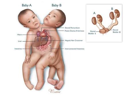
We all know that 3D printing technology inApplications in the Medical FieldIt can be divided intoThree Levels, the closer the application is to the human body, the greater the technical difficulty; conversely, applications farther from the body are relatively simpler and easier to implement. The first layer involves extracorporeal applications. For example, 3D printers can be used toCT、MRgenerating 3D images and models from 2D images, making it more intuitive for physicians to analyze conditions, aiding in preoperative analysis and planning, and reducing surgical risks; Texas Children's Hospital in Houston has implemented this level of technology.
Director of Radiology Research and Cardiac Imaging, Texas Children's Hospital, HoustonRajesh KrishnamurthyA medical doctor stated that skeletal structures or specific organ models can be 3D-printed, and the complex model used in this surgery was one such example. This isRajesh KrishnamurthyThe doctor’s first attempt to use a model to represent the entire infantAnatomy. This model includesSkeletal, cardiovascular, vascular, gastrointestinal, and gynecological structures,So, what exactly is the surgical procedure?
KrishnamurthyAt a press conference during the 2015 annual meeting of the Radiological Society of North America, Dr. [Name] described the surgical procedure:Knatalye HopeandAdeline Faith MataThey are twins, born on April 11, 2014. These two twinsFrom Chest to PelvisConnected together, but with separate brains and hearts.KrishnamurthyThe doctor did not initially intend to create a sensational 3D print. However, the physicians obtained all available information on the conjoined twins and realized that using3D PrintingIt is feasible to integrate all information through technology. Thus, when the twins were approximately five months old, the research team beganModel Imaging. Radiologists used a technique calledProspective ECG Gating Technology in Target ModeBy stabilizing cardiac and pulmonary motion in the images, the research team was able to obtain detailed views of cardiovascular anatomy while maintaining low radiation exposure. Contrast agents were then administered intravenously to each of the conjoined twins separately, with one twin also receiving oral contrast. The model can visualize blood flow in the various organs of the conjoined twins.
The entire model took three weeks to complete, includingDallasIt took the company one week to produce the 3D model. The cost of materials and printing time was approximately $4,000. Different colors and textures were used to represent bones, organs, and blood vessels. Once the model was completed, doctors could remove individual parts to observe deep anatomical structures. On February 17 this year, a team comprising 12 surgeons, six anesthesiologists, and eight nurses successfully completed the 26-hour surgery. Subsequently, both infants showed good postoperative growth.
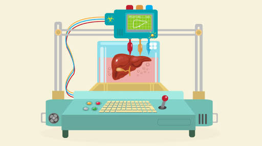
andCleveland Clinic, USAAs one of the world’s busiest and most innovative medical centers, it has seized the prime opportunity to implement 3D printing practices. While other hospitals were still actively introducing this technology, it had already begun large-scale implementation, with its initial application being the 3D printing of liver models.
In fact, as early as 2012, the hospital had already begun researching 3D printing applications in medical practice. A renowned hepatologist from ClevelandNizar ZeinAfter being introduced to and gaining an in-depth understanding of 3D printing in 2012, I began considering the application of this new technology in high-risk liver surgeries. Due to the extremely complex structure of the liver, surgeons may inadvertently sever critical structures such as blood vessels during the procedure. Although advanced medical technologies allow physicians to carefully study three-dimensional computer models of the liver prior to surgery, two-dimensional visual perception still imposes significant limitations on surgical planning. In contrast, tangible 3D-printed liver models offer a distinct advantage by enabling surgeons to conduct preoperativeSurgical Practice, this has become an increasingly common adjunct in surgical procedures.
It is undeniable that 3D printing technology has developed at a remarkable pace,Nizar ZeinBack in 2012, just three years ago, it took approximately six weeks to 3D-print a liver model, whereas now it takes only about 48 hours. This advancement has not only significantly reduced production time but also brought about a qualitative improvement in printing precision. Early printed models were relatively coarse, while current models are far more refined, clearly visualizing the liver’s vasculature and biliary ducts for physicians. Additionally, different anatomical regions can be printed separately. Over the past three years, Nizar has 3D-printed more than 20 liver models, bringing hope and benefit to many patients with liver diseases.
Generally, in terms of the extent of 3D printing applications in healthcare,Second LayerMoving one step closer to the human body, the focus has shifted to printing medical assistive devices. For instance, to achieve greater precision in dental implant procedures, 3D printing technology can be used to create a model of the patient’s teeth. The position, angle, and depth of the implant are first simulated using computer software, and then a “Guide Plate", with the "guide plate," teeth can be implanted very accurately.
Over the next decade, 3D-printed medical models and customizedSurgical GuideIt is highly likely to become the standard procedure for many surgeries, includingCardiac Surgery, Mandibular Surgery, Total Knee Arthroplasty, Hip Replacement, Intracranial Implantation, Shoulder Joint Surgery, Spinal Surgeryor more other surgeries.
On the other side of the straitTaiwan, apparently moving more hastily in the practice of 3D-printed medicine,National Cheng Kung University Douliu Hospitalis one of the hospitals with outstanding 3D medical practices, having successfully completed multiple oral surgery cases using 3D-printed auxiliary tools.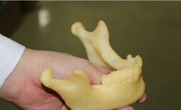
Douliu Cheng Kung University HospitalPreviously completed a case of computer-assisted printing of the mandible.Oral Cancer Reconstruction Surgery, the patient is a 78-year-old male who previously received treatment and follow-up care for oral cancer at another hospital. Due to recurrence of the oral cancer, he sought further medical attention at the Department of Oral and Maxillofacial Surgery, Douliu Cheng Kung University Hospital. Following diagnostic evaluations, including computed tomography (CT), Dr. Chen Meng-Yan, a dental specialist, decided to perform a mandibular resection to completely remove the tumor. To achieve comprehensive reconstruction of the resulting facial defect, a customized 3D-printed mandibular model was prepared preoperatively.
Dr. Chen Mengyan, the lead surgeon, stated that the 3D printing-assisted mandibular reconstruction technique was developed by Dr. Wang Dongyao, Director of the Department of Oral and Maxillofacial Surgery at National Cheng Kung University Hospital, and Professor Fang Jingjing from the Institute of Manufacturing Information and Systems at National Cheng Kung University. Leveraging computer-aided design, this technology enables customized shaping of the jawbone to restore mandibular integrity, thereby recovering both oral function and facial aesthetics. The technique has been implemented at National Cheng Kung University Hospital for ten years and has been adopted by multiple hospitals across Taiwan.
Dr. Chen Mengyan utilized technology locally developed at National Cheng Kung University to reconstruct the patient’s mandibular images prior to surgery. He then customized a 3D model of the jaw and employed 3D printing technology to fabricate a model for the titanium plates to be implanted in the oral cavity. The oral surgery team accurately resected the tumor, followed by mandibular reconstruction using the titanium plates. Subsequently, the plastic surgery team performed meticulous microsurgical free flap reconstruction to repair the defective cheek. This approach not only reduced operative time by 20% but also successfully restored oral function.
ExceptTaiwan,United KingdomNaturally, they are not to be outdone. Some hospitals in the United Kingdom have made equally rapid progress in 3D printing medical practices, continuously exploring more practical applications of 3D printing in medicine. Among these, British doctors have used 3D printing to assist inPediatric Lung LavageSurgery.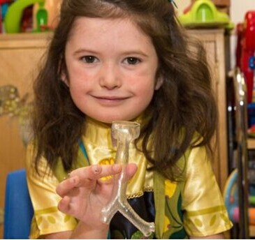
Katie Parkeis an adorable six-year-old girl from Northern Ireland. Unfortunately, she has been diagnosed with a condition known asPulmonary Alveolar Proteinosis(PAP) is a rare disease. This condition leads to significant accumulation of proteins and phospholipids in the alveoli or air sacs of the patient's lungs. Such deposits impair the lungs' ability to absorb oxygen, potentially resulting in dyspnea.
Fortunately, this disease is treatable, but the treatment process is extremely painful—so much so that merely hearing about it sends chills down one’s spine. The most commonly used treatment method is known asWhole Lung Lavage, which involves lung lavage with saline solution. This surgical procedure is highly complex, requiring one lung lobe to remain ventilated while the other is cleaned. Since the size of the bronchial airways varies significantly among individuals, particularly in children, physicians are often compelled to trial tubes of different sizes to identify the optimal fit, thereby consuming valuable time during the operation.
To this end, LondonGreat Ormond StreetThe hospital’s physicians devised a solution to enhance the safety of little Katie’s surgery: they obtained her CT scan data and used it to 3D-print a rubber model of her trachea. This enabled the medical team to determine and prepare all necessary matching instruments prior to the operation. With the corresponding tracheal model prepared in advance, Katie’s surgical time was significantly reduced.
As Katie will undergo multiple such surgical procedures in the future and is still a growing child, the shape and size of her lungs change with each operation. Therefore, doctors need to print an accurate tracheal model before each surgery. Fortunately, this 3D printing process is low-cost and can be completed within a few hours.
It is worth noting that the hospital had already initiated research on using 3D printing to assist in high-risk surgeries. Currently, they also plan to 3D print a series of models for training purposes, enabling physicians to obtain ample practice before performing actual surgeries on patients.
The application of 3D printing in the first two layers does not involveChanges in Human Organs, and thus relatively easy. Upon reaching the third layer, i.e.,Tissues, Stents, Bones, and Organs Implanted in the Human Body...and so on. The application at this level requires a high degree of technical sophistication; while it remains some distance away from widespread clinical adoption, many hospitals are nonetheless stepping forward as pioneers, opening a door to the future of surgical medicine.
Australia, located in the Southern Hemisphere, was the first to complete a 3D-printed case using 3D printing.Titanium Spinal ImplantsSurgery. It is reported that this is the first application of 3D-printed in AustraliaSpinal Implants, patients who received the implant areAmanda GorvinMs.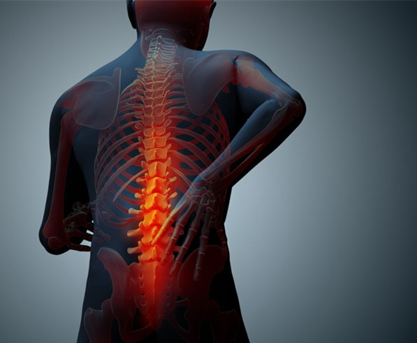
Due to the structural abnormality of its fifth lumbar vertebra and severe degeneration of the adjacent intervertebral disc,GorvinShe had long been plagued by lower back pain. Later, she was referred toNorth Gosford&Prince of WalesHospital Spine Surgery SpecialistMarc CoughlanDr.CoughlanSurgery was considered the optimal solution, but a challenge stood in the way of its implementation. Due to the patient’s unique spinal anatomy, Coughlan was concerned that an off-the-shelf standard implant would be unlikely to alleviate the patient’s symptoms. What was the solution? 3D printing, of course. To this end, the physicians approached a medical device company based in Melbourne.Anatomics, hoping to collaborate forCoughlan3D Printing an Implant.
Anatomics is pleased to announce that, in collaboration with Professor Milan Brandt and his team at the RMIT University Centre for Additive Manufacturing, they have jointly designed and developed a 3D-printedTitanium Alloy Spinal Implants。
An elite team of scientists and professionals from Anatomics and RMIT performed a CT scan of the patient’s spine to create a precise 3D image, which enabled the treatment team to design a customized implant. Another medical device supplier, LifeHealthcare, also provided assistance. The implant took approximately ten hours to 3D print and was subsequently implanted into Gorvin by surgeons.
Three months after the implantation surgery, Gorvin has returned to a normal life and is completely free from the severe pain he previously endured.
Not just Australia, but also in Western EuropeSpainIt is also actively engaged in 3D printing medical practices, with its focus directed towardSternal and Rib Implantation Surgery。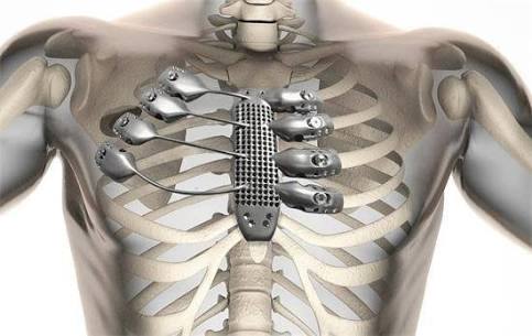
A 54-year-old Spanish man was diagnosed with chest wall sarcoma. Complicating matters, the malignant tumor had grown in a particularly challenging location, fusing with part of his rib cage. Consequently, complete resection of the tumor would require surgeons to remove a portion of his skeletal structure as well. Due to the significant anatomical variability of human ribs and the difficulty in replicating their shape, titanium plates are typically used to reinforce the structural integrity of the rib cage. However, this approach is suboptimal, as it carries risks of loosening and increased complications. Fortunately, 3D printing technology proved to be a timely and effective solution.
Hospital Universitario de Salamanca, Spain's surgical team, based in Melbourne, Australia,AnatomicsA medical device company has custom-engineered a highly complex titanium component for a patient, designed to integrate seamlessly with the patient’s sternum and rib structure.
The team first performed high-resolution CT scans of the patient’s thoracic cavity, then reconstructed a model encompassing the tumor to enable surgeons to plan preoperatively and minimize trauma to the patient’s body, while allowing Anatomics to subsequently fabricate a thoracic implant that fits perfectly.
To ultimately print the implant, Anatomics sent the files to Lab 22, a 3D printing laboratory under Australia’s Commonwealth Scientific and Industrial Research Organisation (CSIRO), where it was fabricated using an Arcam metal 3D printer priced at up to AUD 1.3 million.
Its principle is through powerfulElectron Beamto melt metal powder, then layer by layer to stack out 3D printed objects. Adam Knight of CSIRO said in a blog post: "When you're waiting for surgery to save your life, this is definitely the top priority." Previously, Lab 22 had also helped the manufacturer create a 3D-printedTitanium Root Bone(heel bone) implant. After 3D printing is completed, a polishing and finishing process is still required.
After completion, the finished product was express-delivered to Samarak University Hospital and successfully implanted into the patient’s chest. Two weeks have passed since the surgery; the patient has been discharged and is recovering well.
Over the past two years, numerous successful 3D printing-assisted implant surgeries have emerged, although 3D-printed implants for surgical useRibs and SpineAlthough it still accounts for only a tiny fraction of the overall implant market, it has emerged as another major breakthrough in using 3D technology to treat previously incurable diseases.
The report projects that the total global market revenue for 3D-printed medical implants will reach $3.5 billion by 2020. Some institutions believe that common 3D-printed orthopedic applications have been sufficiently validated, paving the way for broader commercial adoption of medical implants. Meanwhile, the application of 3D-printed medical implants is rapidly expanding to include knee, spinal, shoulder, and other implants, addressing the limitations of traditional treatments in complex cases.
Now, professional medical transplant providers are using industrial printers to create customized implants for patients who would otherwise struggle to find viable options through traditional implantation methods. As the population ages and physiological functions decline in older adults, it becomes increasingly unlikely that they can undergo two or even three surgeries. Manufactured via 3D printingProfessional ImplantsIt is often the only approach with the potential for a durable cure.
In addition toModels, Surgical GuidesandImplantIn addition to established and proven practices, physicians around the world are also actively exploring new, more experimental medical applications with the potential to be life-changing.
So, what comes next?3D Cell Printing!

Cell PrintingThis belongs to a relatively cutting-edge research area and is based onMicrodroplet Depositiontechnology—printing layer by layer with one layer of thermosensitive adhesive material and one layer of cells; the thermosensitive adhesive material degrades after temperature regulation, forming a cell-containingThree-dimensional structure. Cell printing can provide new research tools for life sciences and basic medical research fields such as regenerative medicine, tissue engineering, stem cells, and cancer; offer new clinical medical technologies for constructing and repairing tissues and organs; and promote the development of surgical reconstruction and plastic surgery, regenerative medicine, and transplant medicine. It can be applied to drug screening technologies and controlled drug release technologies, holding broad prospects in the field of drug development. Regarding the application of 3D printing in the medical field, a concept known as the “3D Printing Life Ladder” has been proposed,Inanimate ProstheticsLocated at the bottom tier of the ladder; in the middle is JianSingle Active Tissue, such as bone and cartilage; above the simple tissues will be veins and skin; closest to the top tier of the hierarchy will beComplex and Critical Organs, such as the heart, liver, and brain; while the top tier of the life ladder will be the complete life unit.
Furthermore, research on the application of 3D printing technology in healthcare also involvesNanomedicine, pharmaceuticals, and even organ printing. Manufactured via 3D printingMedical ImplantsIt will improve the quality of life for some people around you, because 3D-printed products can be customized to precisely match individual body shapes. Today, this technology is already being applied to manufacture superior titanium bone implants, prosthetic limbs, and orthotic devices. Experiments on bioprinting soft tissues are currently underway, and it is likely that 3D-printed blood vessels and arteries will soon be used in surgical procedures.
Healthcare is a topic of ongoing discussion today, but not all aspects of healthcare are policy-related. Innovations in treatment and technology are transforming the way people receive medical care, although some methods either struggle to keep pace with the times or fall short of the goals of improving lives or saving them. Now, with the rapid development of the additive manufacturing industry (these increasingly popular technologies are often referred to as 3D printing), the future of the healthcare industry is becoming increasingly clear.