Top 10 Medical Breakthroughs of 2015: Innovations Transforming Healthcare
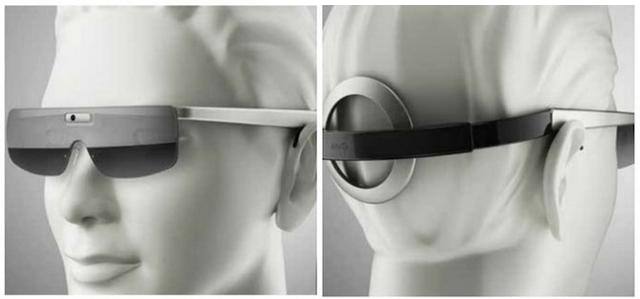
(Electronic Bionic Eye)
2015 has come to an end. The pace of development in medical technology last year was so rapid that it sometimes felt difficult to keep up with the times; indeed, new technological breakthroughs could emerge overnight. Nevertheless, we are eager to present a year-in-review. These cutting-edge innovations from 2015—some of which may have first appeared in 2014 but only gained widespread recognition and began benefiting the public this year; others still in laboratory research yet holding substantial market potential; and some already commercialized, demonstrating remarkable innovation—collectively represent key trends in medical technology. While not exhaustive, this overview highlights significant advancements. Let us begin!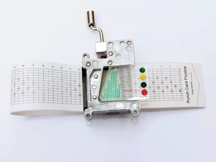
1. Microfluidic Bio-Diagnostic Chip Capable of Diagnosing Multiple Diseases
IBM Researchers Have Developed a Method for the Quantitative Detection of Multiple DiseasesPortable Diagnostic Chip. Due to IBM researchersLuc Gervais and Emmanuel DelamarcheGroundbreaking research demonstrates that capillary tension-driven microfluidics can dramatically transform the landscape of point-of-care diagnostics.
AtIBM Zurich Research LaboratoryScientists have creatively developed a one-step immunoassay by integrating microfluidic components with reagents such as analyte molecules, detection antibodies, and capture antibodies. This sandwich immunoassay can be completed using only 5 microliters of human serum sample, with results read via fluorescence microscopy. The functional microfluidic elements consist of a reaction chamber sealed with a PDMS substrate, a sample collector, a delay valve, a deposition zone for detection antibodies, a capillary pump, and fluidic inlets and outlets.
The chip used in the one-step immunoassay is equipped with meticulously designedMicrofluidic Channel, enabling precise control over the volume and flow rate of the liquid to be analyzed on the chip. The deposition areas for detection antibodies and capture antibodies can be precisely defined, allowing assays to be performed with minimal sample volumes and small detection areas. Moreover, due to the small size of the detection areas, multiple proteins can be analyzed simultaneously on a single chip.
The chip prototype used in this study is capable of detecting the cardiac biomarker C-reactive protein (CRP). Furthermore, the chip provides quantitative detection for cancer, allergies, viruses, and bacteria, and can be applied to diseases associated with any known protein biomarkers.
Further development of multifunctional chips that operate entirely on capillary tension demonstrates the potential of PDMS as a bio-substrate for diagnostic tests and lab-on-a-chip applications.
Coris BioConcept and IBMThey are collaborating to bring a one-step diagnostic chip to market. Researchers hope to eventually manufacture the device using plastic components and promote its adoption in hospitals. Furthermore, IBM plans to leverage its strengths in semiconductor technology and manufacturing expertise to actively collaborate with other biological researchers—including Neuro-Zone, a company developing equipment for detecting brain cells in the treatment of neurological disorders—to co-develop point-of-care products.
2. 3D-Printed Tablets for Epilepsy Treatment
New Generation “Miracle Drug”3D Printing“It is now possible to 3D-print edible items, and even medications! A considerable number of researchers are already dedicated to the development of this technology, such as 《International Journal of Pharmacy》reports that researchers at University College London have successfully trialed 3D-printed tablet technology. In this regard, the U.S.-based company Aprecia is ahead of the curve, having announced that the U.S. Food and Drug Administration (FDA) has approved the market launch of its so-called “world’s first” 3D-printed drug, Spritam (levetiracetam) orally disintegrating tablets, which are used to treat epilepsy.
It must be said that this is a milestone in the entire field of 3D printing, as it enables the simultaneous manufacturing of millions of identical products.
Beyond convenience and personalization, another indispensable advantage of 3D printing technology is its superior structural controllability—a capability unattainable by traditional manufacturing methods. The benefit of 3D-printed medications lies in their highly porous structure, which allows them to dissolve with minimal water in the mouth, thereby facilitating administration for patients with dysphagia, such as those experiencing epileptic seizures. Although conventional methods can also produce porous drug formulations, the process is significantly more challenging and costly.
Looking to the future, 3D-printed drugs may be able to meet more personalized medical needs. Some researchers also believe that 3D-printed drugs could provide a cheaper way to produce medications for developing countries.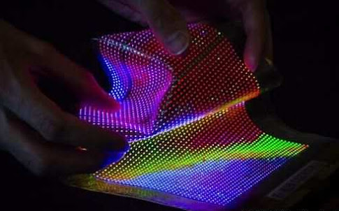
3. Flexible Microelectronics
Flexible Microelectronics (Flexible Microelectronics) can adapt to the irregular shapes of human tissues and move in concert with them, thereby enabling sensing capabilities; in the future, they may even respond to various physiological parameters. Google’s smart contact lens is a representative example in this regard.
Last year, Google announced that it was developing a smart contact lens capable of helping diabetic patients monitor their blood glucose levels by analyzing the glucose content in their tears, thereby sparing them the pain of blood sampling for testing. The contact lens is embedded with tens of thousands of microtransistors and an antenna as thin as a human hair, enabling wireless transmission of data to mobile devices such as smartphones.
This year, John Rogers, a professor at the University of Illinois, collaborated with a research team from Washington University to develop a flexible cuff that can wrap around the exterior of a beating rabbit heart to monitor its electrical activity in 3D. In the near future, this technology may be used for highly precise detection and response to cardiac arrhythmias.
Furthermore, Professor Rutgers has also developed a flexible skin “Patch”, capable of recording electrocardiogram (ECG) and electroencephalogram (EEG) signals, and transmitting them wirelessly to smartphones or other devices.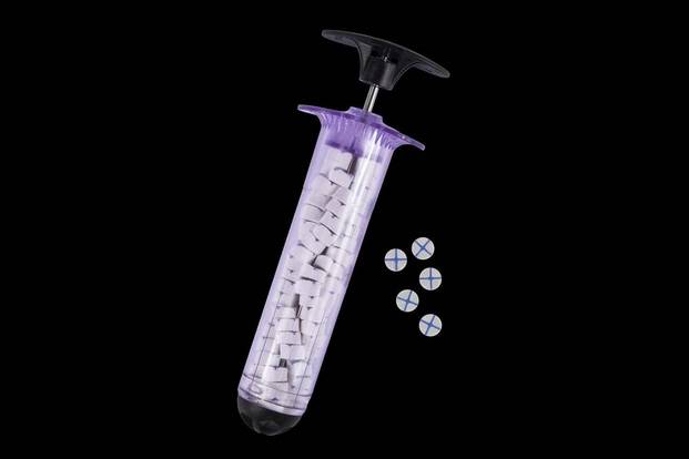
4. Rapid Hemostasis System
In the past, when soldiers were unfortunately shot on the battlefield, the rudimentary nature of on-site emergency medical care was hardly any better than the gunshot wound itself. Gauze had to be immediately packed into the gushing, bleeding wound. Sometimes, the temporary cavity formed by the bullet penetrated as deep as five inches. Faced with such severe injuries, limited first-aid measures proved largely ineffective; despite exerting tremendous effort to pack gauze into the wound, hemostasis often failed. Medics were then forced to remove the gauze and repeat the entire procedure. For the wounded, this experience was truly devastating, causing both profound psychological trauma and physical collapse.
Former U.S. Army physician John Steinbaugh said, “Before we provide aid, we must first kick their guns away; otherwise, as soon as we move in…”
No matter how hard medical officers tried, many wounded soldiers could only watch helplessly as they bled to death; severe hemorrhage is the leading cause of death among battlefield casualties. Dr. Steinbaugh, a military physician who served for many years in Iraq and Afghanistan, noted that gauze is essentially useless when it comes to serious injuries. After retiring in April 2012 due to a head injury, Dr. Steinbaugh joined RevMedx, an Oregon-based startup, where he collaborated with a team of veterans, scientists, and engineers to develop methods for rapid hemostasis.
In April 2014, the U.S. FDA approved their new invention:XStat, a portable, modified high-pressure syringe that delivers a large volume ofCoated SpongeInjected into the wound. XStat can save the lives of many soldiers on the battlefield, alleviate the suffering caused by crude first aid, fill wounds more rapidly than gauze, and achieve rapid hemostasis.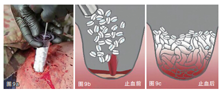
This product acts with remarkable speed: within just 15 seconds, it completely seals the wound and generates sufficient pressure to halt profuse bleeding. Furthermore, because the hemostatic sponge firmly adheres to the entire wound site, it will not be expelled even from wounds with severe, gushing hemorrhage.
However, inserting the hemostatic pellets into a wound requires considerable skill. On the battlefield, medics must carry various life-saving equipment, dash across the field under heavy fire, and even bear arms to engage in combat. Therefore, RevMedx needed to develop a device that was sufficiently portable and lightweight to deliver hemostatic sponges deep into wounds. The development team designed a polycarbonate syringe with a diameter of 30 mm, with the hemostatic sponge stored directly inside the syringe to save space. To use it, the plunger is pulled back, the syringe is inserted into the wound as close as possible to the bleeding vessel, and the medication is administered promptly—before the wounded soldier resorts to shooting himself.
Writing this, I suddenly thought of a joke:
Soldier A: "I'm so depressed; I got bitten by a venomous snake."
Soldier B: Don't panic. I have hemostatic sponge. Come on, apply it quickly.
Passerby A dies.
5. Luminescent Eye Drops for Early Detection of Glaucoma
Glaucoma is one of the three leading causes of blindness worldwide. It is an ocular condition characterized by intermittent or sustained elevation of intraocular pressure. Persistent high intraocular pressure can damage various ocular tissues and impair visual function; if left untreated, it may lead to complete loss of visual field and ultimately blindness. However, glaucoma is typically asymptomatic in its early stages and often goes undetected, being identified only during routine eye examinations. As a result, patients frequently do not become aware of visual impairment until a decade after disease onset.
A new testing method capable of detecting in the eye is being developed through a collaboration between University College London, the Wellcome Trust, and Imperial College Healthcare NHS Foundation Trust.Dead Neurons, detect glaucoma ten years in advance, and hold the promise of saving the vision of millions.
The research team used a natural protein containing dye (annexin) to detect dead cells. When encountering dead nerve cells, the compound fluoresces. Fluorescence detection can be performed using traditional eye examination equipment. If a patient has a large number of dead cells, it indicates a high risk of glaucoma, and drug treatment is recommended in an effort to stop nerve cell death before vision loss occurs.
The research team is currently developing this dye into an eye drop formulation. Meanwhile, researchers believe that this approach could potentially be applied to the early diagnosis of other neurological disorders, such as Alzheimer’s disease and Parkinson’s disease.
Upon the successful development and market launch of this product, it is expected to be widely welcomed. This technology has the potential to restore vision for millions, reduce blindness rates, and bring light back to the world. Furthermore, its applications extend broadly into the field of chronic neurological diseases; through early detection and prevention, the dream of “immortality” may no longer be out of reach.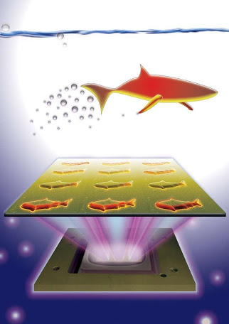
6. 3D-Printed Microrobotic Fish
As is well known, fish meat is delicious, rich in protein, vitamins, iron, and other nutrients, with high nutritional value and easy absorption, making it excellent for health. What about fish-shaped robots?
Researchers at the University of California, San Diego have developed micro-robots capable of swimming in liquids and serving multiple purposes, utilizing new 3D printing technology. This type of robot is known as “Microfish” robots, which can be driven and controlled through hydrogen peroxide chemical reactions and magnetic forces, enabling multiple functions such as detoxification, remote sensing, and targeted drug delivery.
The currently printed robotic fish adopt a self-propelled, magnetically actuated design. Researchers stated that they employed a technique known as micro-continuous digital light processing (DLP) high-resolution 3D printing. This process enables the researchers to print hundreds of miniature robotic fish, each 120 micrometers in length and 30 micrometers in thickness, in a single batch. By using computer-aided design (CAD) software, they can rapidly modify designs to fabricate robotic fish with appearances resembling sharks, manta rays, or even birds.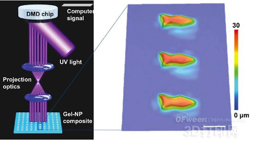
Nanoengineers can conveniently incorporate nanoparticles with different functions into “Microfish”A specific part of the body: Platinum nanoparticles are printed at the fish tail, where they undergo a chemical reaction with hydrogen peroxide, thereby serving as fuel to propel it.“Microfish“Forward movement; by printing iron oxide nanoparticles on the head of the fish, its steering can be controlled via the magnetic properties of iron oxide. By altering the shape of the ‘microfish’ and adjusting the amount of hydrogen peroxide used, researchers were able to control the swimming speed of the ‘microfish’ in liquid.”
The polydopamine nanoparticles within the robotic fish can neutralize toxins. As the neutralization process progresses, the robotic fish emits increasingly intense red fluorescence, with the glowing red signal becoming denser. This indicates that the robotic fish serves a dual function as both a detoxification agent and a toxin sensor. Researchers also believe that these robotic fish can be utilized for targeted drug delivery, environmental protection, and many other applications.
Simple “microfish” can not only serve as future “Medication Courier" or "Scavenger,” and can also assume various shapes, which may mean that one day in the future, your body could become a “zoo.”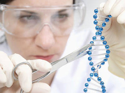
7. CRISPR Gene Scissors
Since the beginning of this year, scientists have usedCRISPR SystemRewriting the Equation of Life: This novel gene-editing tool can rewrite DNA, ushering us into an era where diseases can not only be prevented but also eradicated, where plant and animal genomes can be edited, and even “designer babies” (via embryonic gene modification) become possible.
The CRISPR-Cas9 system, born at the Massachusetts Institute of Technology, is essentiallyGenome Search and Replace Tool. Don’t want DNA sequences associated with a specific disease? The Cas9 protein can cut out or even replace them.
Jennifer Doudna, a biologist and co-discoverer of CRISPR, revealed that we now possess a molecular scalpel capable of cutting the genome. All previous technologies were somewhat akin to sledgehammers… This discovery has provided scientists with a practical tool, which is incredible. Dustin Rubinstein, head of the CRISPR Collaborative Laboratory at the University of Wisconsin–Madison, told us that gene editing can transform issues in cancer research and neuroscience into problems of chemical engineering and even energy production.
From now on, you are limited only by your imagination.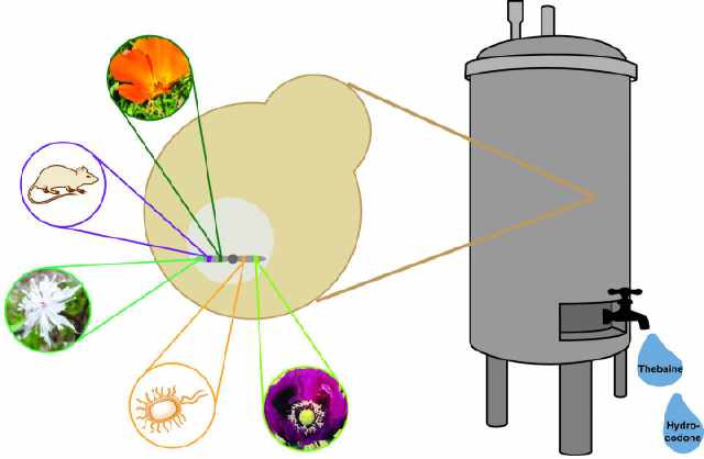
8. Synthesizing Opioid Analgesics Using Yeast
Many drugs are derived from plants. Our ancestors chewed leaves or brewed them as tea, and later used chemical methods to extract and concentrate their active ingredients into refined pills. For thousands of years, yeast was used exclusively for fermenting wine, brewing beer, and leavening bread, with little application in biopharmaceutical contexts. Now, researchers at Stanford University in the United States have genetically engineered yeast to produce opioid painkillers. This breakthrough suggests that many different types of plant-derived pharmaceuticals can be produced using a faster and potentially more cost-effective method.
This composite biological feat was published in an issue of Science magazine this year: by introducing 21 genes from plants, bacteria, and rodents, a “Drug Production Line”, which can convert sugar step by step into thebaine—the precursor of morphine. The research team also found that further engineered yeast can produce hydrocodone, a widely used analgesic chemically synthesized from thebaine.
Synthetic biology is a fascinating field in which scientists view the genes of various organisms as components and design them like “Circuit”assemble them in the same way to accomplish various tasks envisioned by the designers.
Previously, synthetic biologists designed a [organism/system] capable of producingArtemisinin(antimalarial drugs) in yeast, but this only required the insertion of a few plant genes. This time, however, is far more complex; producing thebaine requires the introduction of 21 genes from different species, while producing hydrocodone requires even more, with 23 genes.
Ultimately, by engineering the metabolic pathways of yeast, synthetic biologists have successfully synthesized opioid analgesics. This process is akin to homebrewing beer, suggesting that more pharmaceuticals could be manufactured using similar methods in the future.
In this paper, the authors acknowledge that this new manufacturing process for opioid analgesics may heighten concerns regarding “the potential issues of opioid misuse.” Given the widespread use of opioids in the United States, the focus is on the potential for misuse.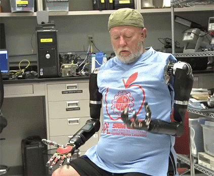
9. Smart Sensing Limbs
Imperial College London Unveils Novel Robotic HandImperial College London has recently unveiled a novel robotic hand equipped with sensors that directly detect minute vibrations in the arm’s muscle fibers, enabling users to effortlessly control the device through simple muscle responses and arm movements. This technology holds promise for the future development of more advanced and cost-effective mechanical prosthetics for individuals with disabilities.
Researchers involved in the project explained that most previous mechanical prostheses were controlled by electrical signals generated by muscle activity. This approach requires sensors to be in contact with the user’s residual limb to detect these signals. However, such electrical signals are highly susceptible to interference; for instance, perspiration can disrupt signal transmission, thereby impairing the control of the mechanical prosthesis. Furthermore, the relatively high costs associated with the manufacturing, debugging, and calibration of these devices hinder their widespread adoption.
Furthermore, the researchers also equipped the robotic arm with aMotion Sensor, the control modes of the robotic hand can be further refined. Users can control the robotic hand to pick up objects of various sizes through a series of simple muscle responses and arm movements. An amputee volunteer has conducted preliminary trials with this robotic hand and reported satisfactory results.
Similarly, researchers at the École Polytechnique Fédérale de Lausanne (EPFL) in Switzerland and the Sant’Anna School of Advanced Studies (SSSA) in Italy have developed a novel smart prosthetic limb equipped with tactile sensors at the fingertips to perceive touched objects. By connecting to the user’s nervous system, the prosthesis provides feedback on information such as object stiffness and shape, thereby simulating human hand sensation to a certain extent.
These R&D achievements will make mechanical prosthetics more stable, flexible, and easier to control. In the future, researchers will further enhance the stability of this prototype robotic hand, helping individuals with disabilities better control their prosthetic limbs.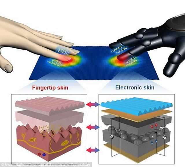
10. Artificial Plastic Skin
A newly developed ultra-sensitive electronic skin from the Ulsan National Institute of Science and Technology in South Korea is capable of simultaneously detectingHeat and Temperaturechanges.
This latest electronic skin was developed by Professor Jonghwa Park and his colleagues at the Ulsan National Institute of Science and Technology (UNIST) in South Korea. Human skin contains unique microscopic structures of the epidermis and dermis, as well as sensory receptors. The miniature ridge-like protrusions on the fingertips are designed to fine-tune the perception of surface textures and transmit sensory information to the brain.
In tests, the grooves of the electronic skin were able to sense water droplets flowing across them and detect the pressure exerted by a strand of hair placed on its surface. Existing electronic skin technologies enable robots and mechanical prosthetics to grasp and manipulate objects, identify surface texture and hardness, and perceive the temperature of objects. However, prior to this development, it was difficult to achieve highly sensitive simultaneous detection of heat and different types of pressure with electronic skin.
Researchers tested the electronic skin's response to sensory changes using water droplets and found that it could detect falling droplets under varying pressure and temperature conditions. Meanwhile, they discovered that the artificial fingertip skin could detect the minute pressure generated by a single human hair.
When the electronic skin is attached to the human wrist, the wrist blood vessels expand and contract. The electronic skin can be used to monitor pulse pressure by detecting changes in skin temperature.
As early as September this year, researchers from Stanford University in the United States developed a sensitive tactile artificial skin that can not only detect pressure but also transmit signals to nerve cells.
This technology operates on principles similar to the iPhone’s fingerprint recognition system and holds promise for replacing skin damaged by burns or used in prosthetic limbs, enabling patients to genuinely experience a sense of touch.
Professor at Stanford UniversityZhenan BaoHe is the principal investigator of this project. The team mimics human skin receptors by incorporating sensors into artificial skin. These sensors are capable of collecting dynamic pressure data, and researchers hope to one day “transmit” this data to the brain in some form.
Although electronic skin has only achieved some breakthroughs in experimental settings, researchers hope that this proof-of-concept study will revolutionize artificial prosthetics by enabling wearers to perceive different surface textures and distinguish between hot and cold temperature changes. This two-layer “electronic skin” features an elastic top layer capable of sensing pressure, while the bottom layer generates biochemical signals suitable for transmission to nerve cells.
If further commercialized, we can use this technology to create more realisticProsthesis, or enhanceWearable Sensorsprecision and medical diagnostic equipment.
Humanoid diagnostic and treatment robots, intelligent electronic scalpel iKnife, transparent mice for experimental use, nanorobots, nanopore sequencers, artificial bloodWait, there are still many medical achievements from this year that have not been mentioned, but this does not mean they are unimportant. In the long river of medical technology development, no matter how advanced the technology is, the most important thing is still us humans ourselves.