AI-Powered Cardiac Imaging Breakthrough: Arterys' Cardio DL Gains FDA Approval and Survival Prediction Algorithms Show 80% Accuracy in Clinical Studies
Recently, two news stories regarding artificial intelligence in the field of disease diagnosis have focused on the diagnosis of heart disease patients. One report highlighted that Arterys, a U.S.-based company, received FDA approval for its Cardio DL, an AI-powered medical imaging analysis system for cardiac MRI. This marks the first AI analysis software for cardiac magnetic resonance imaging to gain regulatory approval. The other report noted that British scientists published an article in the journal *Diagnostic and Interventional Imaging*, suggesting that artificial intelligence can analyze cardiac MRI scans and blood test results from heart disease patients to predict their time of death.
Currently, the most significant advantage of artificial intelligence (AI) in the healthcare sector lies in medical image analysis. According to VCBeat, over 90% of healthcare data originates from medical imaging, yet the majority of this data still requires manual analysis. The drawbacks of manual analysis are evident: first, it lacks precision, as judgments rely heavily on individual experience, leading to a high risk of misdiagnosis; second, there is a substantial supply-demand gap. Data from VCBeat’s VBInsight indicates that the annual growth rate of medical imaging data in China is approximately 30%, whereas the annual growth rate of radiologists is only about 4.1%, resulting in a disparity of 23.9%. The number of radiologists is growing far too slowly to keep pace with the surge in imaging data. In contrast, after undergoing training, AI demonstrates significant advantages over radiologists in both the speed and accuracy of medical image interpretation.
Previously, the most common applications of artificial intelligence in medical imaging that we encountered were primarily for tumor detection and tuberculosis diagnosis. Today, we introduce the use of artificial intelligence in diagnosing heart disease.
FDA Approves First AI Software for Cardiac MRI Imaging
Arterys is a medical imaging company that leverages cloud computing technology for the precise diagnosis of cardiovascular diseases. Its software integrates with magnetic resonance imaging (MRI) scanners. After a ten-minute scan, patient data is transmitted to a HIPAA-compliant cloud server, where it is analyzed via a web browser to deliver accurate diagnostic results. The Cardio DL cardiac MRI analysis system can be used in the management of various cardiovascular conditions, including congenital heart disease, aortic disorders, and valvular heart disease. It automatically captures data on the endocardial and epicardial contours of the ventricles and provides accurate calculations of ventricular function. With high precision and rapid processing, the system completes image analysis in just 10 seconds, significantly faster than manual interpretation by clinicians.
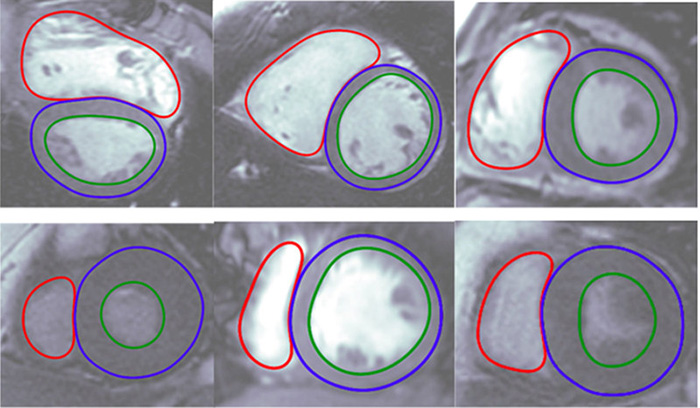
These images display the automatically generated endocardial and epicardial contours by the software.
It is reported that this AI-powered cardiac MRI medical imaging analysis system has not only received FDA 510(k) clearance but also obtained CE certification and approval in Europe, marking its authorization for clinical use. Unlike traditional medical imaging software, Cardio DL leverages deep learning technology. As an increasing number of physicians adopt Cardio DL, Arterys’ cloud platform will enable the software to collect and analyze large volumes of cardiovascular data acquired from MR scanners in real time, thereby accelerating diagnostic decision-making.
“Arterys is dedicated to accelerating data-driven medical transformation through advanced cloud-based medical imaging analysis applications, while safeguarding patient data privacy,” said CEO Fabien Beckers in a statement. “The FDA clearance of this software today marks a significant regulatory milestone. This application demonstrates the power of deep learning, combined with cloud supercomputing technology, to help physicians enhance their interpretation of medical images with both accuracy and automation, thereby alleviating radiologists from burdensome workloads.”
Currently, Arterys will integrate this software into the ViosWorks software system included with GE’s MRI scanners.
AI Can Predict Time of Death in Heart Disease Patients
Recently, British scientists published an article in the journal *Radiology*, with research results suggesting that artificial intelligence can predict when heart disease patients will die.
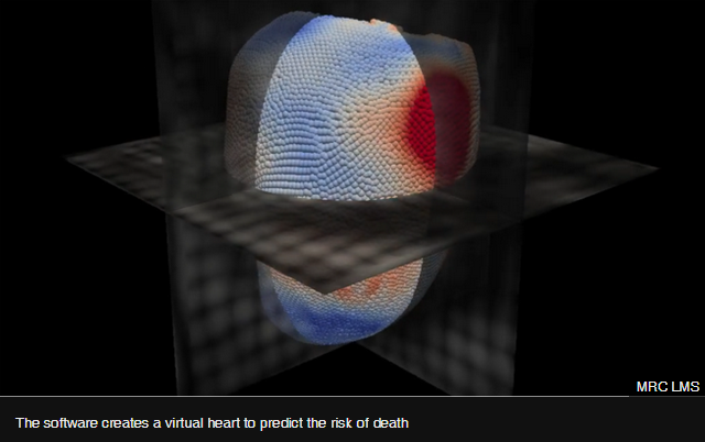
The MRC London Institute of Medical Sciences, under the UK Medical Research Council, states that artificial intelligence software can detect signs of impending heart failure by analyzing blood test results and cardiac scans.
Researchers obtained the above results through studies on patients with pulmonary hypertension. This technology enables physicians to identify patients who require more intensive intervention, thereby saving more lives.
Elevated pulmonary arterial pressure can damage parts of the heart, and approximately one-third of patients die within five years after diagnosis. Current treatment options primarily include direct intravascular drug injection and lung transplantation. However, physicians need to determine patient life expectancy to select the appropriate therapeutic regimen.
Researchers input cardiac magnetic resonance imaging (MRI) scans and blood test results from 256 heart disease patients into an artificial intelligence (AI) software program. The software measured the motion of 30,000 marked points on the cardiac structure during each heartbeat. By integrating this data with the patients’ health records over an eight-year period, the AI software was able to predict which abnormalities would lead to patient mortality.
AI software can predict five-year survival outcomes, achieving an accuracy of approximately 80% in predicting a one-year life expectancy for patients, compared to a 60% accuracy rate for physicians.
One of the researchers, Dr. Declan O’Regan, stated, “Artificial intelligence can help patients develop personalized, optimal treatment plans. The predictive outcomes are highly accurate, assisting physicians in formulating the correct treatment strategies for their patients.”
The research team stated that the technology will next enter hospitals for testing.