AI-Powered Skin Cancer Diagnosis via Smartphone Achieves 91% Accuracy, Matching Dermatologists
Recently, Nature published an article on its cover:Deep Learning Algorithms Achieve 91% Accuracy in Skin Cancer Diagnosis, Rivaling Physicians
I wonder if everyone still remembers how Google’s neural networks distinguish between cats and dogs. Unlike humans, a child can recognize what a cat looks like after seeing just a few examples. In contrast, for a machine to identify a cat, it requires training on tens of thousands of images and must undergo deep learning processes.
Similarly, if high-quality images of skin cancer are provided to an artificial intelligence system, the system can learn to identify skin cancer through machine learning. Stanford University recently published relevant research findings in Nature, comparing this AI system with 24 senior dermatologists and finding that the system achieved an accuracy rate of approximately 91%.
“The first author of the article, Andre Esteva, a graduate student at Stanford University, said: ‘We developed a highly powerful artificial intelligence algorithm capable of learning from data. By writing code, we enabled the system to independently discover what features to identify and seek out.’”
This algorithm is calledConvolutional Neural Network, it first emerged in Google Brain, leveraging its remarkable computational power to enhance the decision-making capabilities of algorithms. Following research at Stanford University, neural networks have been able to identify 1.28 million images across approximately 1,000 different categories; however, researchers need to understand malignant tumors from benign seborrheic keratosis.
Distinguishing dogs from a group of Persian cats may tolerate minor inaccuracies, but differentiating various dermatological lesions to identify skin cancer is a life-critical task that demands extremely high accuracy.
Colorful spots on the skin pose a significant challenge, and distinguishing them algorithmically is a difficult task.
Screening Image Data
Brett Kuprel, a Stanford graduate student and co-author of the article, stated, “Another challenge of the study was that, at the time,”There is insufficient high-quality skin cancer image data of adequate scale to train artificial intelligence algorithms., we must resolve it ourselves.” Even before processing the images, they had to perform some translation work. “We collected a number of images from the internet, collaborated with medical schools to categorize and organize these images, and annotated them with labels—including those in German, Arabic, Latin, and other languages.”
Not only is translation and organization required, but image processing is also necessary.Dermatologists frequently use an instrument called a dermoscope to carefully examine patients; therefore, medical professionals generally diagnose diseases based on medical images with largely consistent magnification and viewing angles. However, images available on the internet vary widely: some are captured with mobile phones, others with specialized instruments or cameras. Furthermore, differences in environment lead to varying image quality, with significant variations in angle, focal length, and illumination.
Finally, the researchers collected approximately 130,000 images of skin lesions, encompassing more than 2,000 distinct diseases. They used these images to create an image library and fed them into the algorithm as raw pixels, with each pixel labeled with additional data describing the associated disease. The researchers then developed a set of algorithms that enabled the system to discern the intrinsic relationships within these images:That is, the rules followed in appearance by disease as it spreads through tissues.
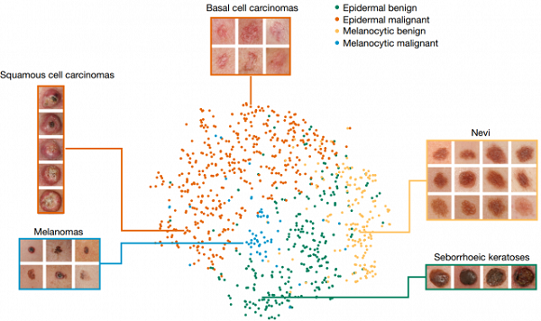
How AI Segments Images of Different Categories of Skin Diseases It Observes
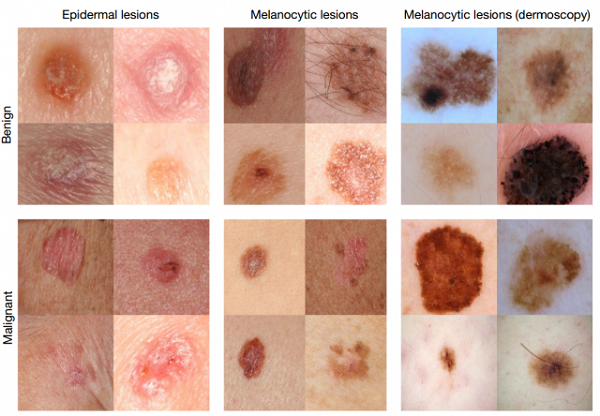
After the research results were released, to verify the accuracy of the algorithm, researchers invited 21 dermatologists from Stanford University School of Medicine to evaluate the algorithm from three perspectives:Classification of Keratinocyte Carcinomas, Classification of Melanocytic Tumors, and Dermoscopic Classification of Melanoma。
In the final test, researchers used only high-quality images of malignant melanomas and malignancies with verified activity, labeling them as requiring treatment, biopsy, or merely reassurance. When the researchers compared the diagnostic results obtained from the artificial intelligence system with those of 21 physicians, they found thatIt performed well in both detecting all cancerous lesions and avoiding false-positive results, achieving an accuracy rate of 91%, which is comparable to the performance of physicians.
Diagnosis Using Mobile Phones
Esteva stated, “Although the team has not yet launched a live app, this has met our expectations. Our original intention was to enable the public to access higher-quality and more convenient medical services.”What excites me even more is that smartphones are now ubiquitous, with each device equipped with a variety of sensors and cameras. We can leverage AI systems to directly analyze smartphone images for skin cancer detection. Moreover, if we can solve the problem of skin cancer, how far behind can other diseases be?”
"In any case, before commercialization, further testing is required, and the algorithm needs to be refined."What matters is that we understand how artificial intelligence makes decisions to distinguish between images.“Advances in computer-aided classification of benign and malignant skin lesions can help dermatologists improve their ability to diagnose challenging lesions and provide better management plans for patients,” said Susan Swetter, Professor of Dermatology at Stanford University and author of the paper. “However, rigorous prospective validation of the algorithms is necessary before implementation in clinical practice.”
Source: https://www.extremetech.com/category/extreme