The Evolution and Future of AI in Medical Imaging: Insights from Tao Xiaodong's Speech at CCF-GAIR
This article is based on Tao Xiaodong’s speech at the CCF-GAIR (Global Conference on Artificial Intelligence and Robotics), compiled and edited by VCBeat.
The presentation is divided into three parts: first, the broader context of artificial intelligence; second, how medical imaging addresses clinical problems; and third, the future directions for medical imaging in the age of artificial intelligence.
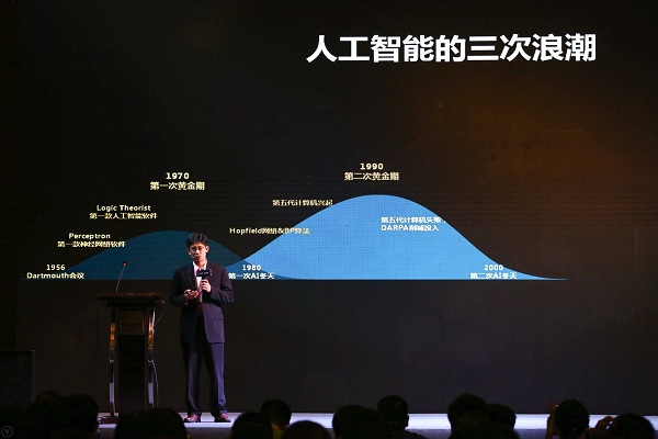
The Three Peaks and Two Winters of Artificial Intelligence
Reviewing the Three Waves of Artificial Intelligence: Since 1956, AI has experienced three peaks and two winters, and it is currently in the third peak.
The first peak stemmed from the introduction of the Perceptron, which addressed several problems that appeared exceedingly difficult in 1956 and 1957. In the aftermath, however, many technologies failed to materialize due to limitations in computational power and various data constraints, leading artificial intelligence into its first winter around 1980.
The advent of the second wave was driven by the introduction of Hopfield networks and the backpropagation (BP) algorithm, along with significant advancements in computational power and storage capacity. However, around the 1990s, the failure of Japan’s Fifth Generation Computer Systems project plunged the industry into a second AI winter.
We are now in the third wave, which originated with the concept of deep learning proposed in 2009. Deep learning has spawned numerous extended algorithms that have been successfully applied across various fields, including the recently popular AlphaGo. These successes have led many to believe that artificial intelligence is truly poised to transform our world.
Three Key Elements of AI Applications: Data, Industry Experts, and Technology
Based on the development of the past “AI+” era, three elements are indispensable for the success of artificial intelligence:Standard Data, Core Technologies, Industry Experts.
First,Standard Data, from our perspective, smart healthcare is inseparable from industry big data. Data must be standardized, complete, and accurate, and it must be relevant to the application.
Currently, the collection, utilization, and representation of big data in the industry present significant challenges. From a big data perspective, these datasets are characterized by poor structure and low homogeneity. Furthermore, given the massive volume of data being continuously collected by our systems, how to effectively represent, utilize, and standardize such data remains a major challenge.
Second,Industry Expert, we require substantial support from industry experts to guide the application of artificial intelligence in specific domains. Furthermore, under expert guidance, we need to establish a knowledge base, clearly distinguishing between knowledge that can be expressed in a machine-readable format and knowledge that cannot. Additionally, after the development of applications, we need experts to assist in validation.
Third,Core TechnologiesCore technology is an area of our particular focus. As technology developers, we must prioritize creating solutions that are more intelligent, practical, and equipped with user retention mechanisms. We aim to minimize disruptions to the workflows of industry experts and physicians, ensuring that artificial intelligence genuinely helps doctors enhance their efficiency and professional performance.
With comprehensive data, support from industry experts, and advanced core technologies, we can leverage artificial intelligence to assist government agencies in policy-making and decision-making, and help hospitals with operational management. AI can also enhance the efficiency of specialist physicians, improve the skills and experience of primary care providers, and support patients in health management. This represents iFlytek’s practical summary of AI applications in smart healthcare.
How Medical Imaging Addresses Clinical Problems
Modern medicine is founded on evidence-based medicine, with medical imaging serving as a critical diagnostic basis. Consequently, there is substantial demand from clinicians for imaging services, including various quantitative analyses and comparisons with historical images.
What iFlytek aims to do is to relay this portion of imaging requirements to engineering technicians, enabling them to present better data information to physicians. Throughout the entire processClinicians, radiologists, and engineering technicians represent three distinct tiers with differing areas of expertise. A critical aspect of ensuring that technology effectively serves clinical applications lies in translating clinicians’ needs into imaging requirements, and subsequently converting those imaging requirements into engineering specifications during communication.。
For instance, if we need to assess the degree of coronary artery stenosis, this will determine the treatment approach—whether stent implantation is required or if management with medication and lifestyle modifications is appropriate.
In this context, the imaging requirements demand clear visualization of the coronary arteries, highlighting the contrast between plaques and blood vessels. This allows for assessment of plaque burden within the vessel lumen, thereby guiding the selection of appropriate therapeutic interventions.
Translating imaging requirements into technical specifications necessitates high-resolution CT imaging. After addressing the imaging challenges, coronary artery segmentation is performed, followed by plaque detection. The resulting data is then integrated with the Radiology Information System (RIS) to ensure accessibility for all relevant stakeholders.
What Problems Should Intelligent Imaging Address? As technical professionals, we often reflect on this in the laboratory. We have frequently discovered that a sophisticated algorithm, though impressive from a technical standpoint, proves entirely useless when presented to clinicians. After extensive communication and collaboration, we have concluded that intelligent imaging technologies—including pattern recognition and image analysis—should fundamentally address three categories of problems:
Ito better present this information to physicians. As medical imaging becomes increasingly accessible and offers higher resolution, physicians are required to review a growing volume of images. However, what clinicians need is not raw data, but actionable information. How can this information be presented more effectively to physicians? This is the core challenge that intelligent imaging aims to address.
IIis to assist physicians in quantitative analysis. Physicians excel at qualitative analysis. Upon viewing the imaging scans, they can roughly identify the issue within one second; however, physicians require tools to make more precise judgments and perform quantitative analysis, which is difficult to achieve with the naked eye alone.
This work encompasses a variety of multimodal analyses, including comparison of historical images and analysis of patient populations, which cannot be performed simply by visual inspection. It requires image segmentation, image registration, and functional image analysis.
Third,Imaging and Intelligent Image Recognition Challenges to Be Addressed in Smart Medical Imaging. These two steps were separated many years ago, with technicians capturing images and physicians performing analysis. In reality, only by integrating both can the system be optimized more effectively to help physicians deliver efficient services.
Take medical imaging as an example. On one hand, there is a shortage of highly skilled technologists, particularly in primary care hospitals, leading to redundant scans and wastage of imaging resources. On the other hand, advanced imaging functionalities are complex; minor adjustments to sequences and parameters by technologists can significantly impact image quality. Therefore, it is essential to establish standardized yet personalized procedural protocols.
StandardizationIt refers to the requirement that the obtained images must have standardized quality.PersonalizationImaging parameters and radiation doses are adjusted based on individual patient characteristics, such as variations in BMI, during CT scans. This personalized approach ensures standardized image quality. Intelligent tools assist physicians and technologists in selecting optimal imaging parameters, ultimately enabling less experienced clinicians at primary care facilities to obtain medical images comparable in quality to those produced by top-tier tertiary hospitals.
Furthermore, with the advent of 3D imaging technology, physicians can sometimes bypass the need to review hundreds of axial CT slices and instead directly visualize volumetric images.
Returning to image analysis, the vast majority of scans reviewed by physicians are from healthy individuals, which constitutes an inefficient use of their time. The current system assists physicians by first filtering out these normal cases and presenting only the remaining images for clinical review.
For abnormal images, the system directly annotates lesions, nodules, and other abnormalities, allowing physicians to simply review the results, thereby improving their efficiency.
Overall, combining imaging with image analysis to screen images of healthy individuals, present abnormal images to physicians, and provide preliminary annotations can improve physicians' efficiency.
Imaging information needs to be combined with clinical information.
The clinical diagnostic process for imaging is as follows:Clinicians determine whether imaging is necessary for patients based on clinical information, medical history, and examination and test results. Imaging requests are then submitted to the Radiology Department, where technologists acquire various images using imaging modalities, and radiologists issue imaging reports. Subsequently, radiologists and clinicians integrate imaging findings with clinical data to jointly produce a clinical diagnostic report that guides patient treatment.。
From this process, we can see that imaging reports alone are insufficient. Clinical information, such as patient history, electrocardiogram (ECG) results, and body temperature, all influence subsequent diagnostic reports. Therefore, iFlytek is now aiming to integrate clinical data with imaging data.
Artificial intelligence has undergone three stages: from computational intelligence to perceptual intelligence, and then to cognitive intelligence. Computational intelligence surpassed human capabilities long ago, while perceptual intelligence can detect many things invisible to the human eye. Cognitive intelligence, which is the focus of contemporary AI discourse, refers to the ability of AI systems to think and reason based on data.
Overall, in the era of artificial intelligence, medical imaging equipment has become easier to operate. The learning paradigm is no longer based on simple, discrete training data, but rather on a comprehensive system of medical knowledge. In clinical practice, imaging information must be integrated with clinical data, and its application requires close collaboration with experts to address real-world clinical problems from a practical standpoint.