Insightec AI Achieves 30-Second Lung and Breast Cancer Detection with 91%–99% Accuracy, Files IPO Prospectus
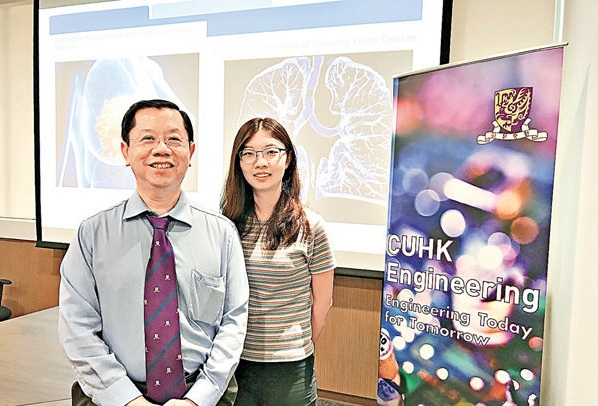
Wang Ping’an (left) and his research team successfully applied AI-based image recognition technology to improve the diagnostic accuracy of lung cancer and breast cancer imaging.
Lung cancer and breast cancer are prevalent high-risk, intractable diseases in Hong Kong. To enhance the efficiency of clinical diagnosis, a research team from the Faculty of Engineering at The Chinese University of Hong Kong has employed artificial intelligence-based image recognition technology. By utilizing deep learning systems to interpret medical images such as computed tomography (CT) scans and histopathological slides, the team conducted research on these two types of cancer. The results demonstrated that the accuracy rates for AI-assisted interpretation of cancer-related medical images reached 91% and 99%, respectively. Moreover, the identification process was accelerated to between 30 seconds and 5 minutes, thereby reducing the rate of misdiagnosis. The team plans to collaborate with local public hospitals, aiming for widespread application of this technology within the next one to two years.
Lung cancer is the leading cause of cancer-related mortality in Hong Kong, with thousands of new cases diagnosed annually. In its early stages, lung cancer often presents as small pulmonary nodules, which appear as tiny, mass-like opacities on lung imaging. Currently, physicians primarily rely on computed tomography (CT) scans of the chest to detect whether patients have small pulmonary nodules.
However, each examination can generate hundreds of CT images. Under normal circumstances, if each image is reviewed visually at a rate of 3 seconds per image, the process would take at least 5 minutes, consuming significant time and energy. Moreover, diagnostic accuracy may vary depending on the physician’s experience and mental state.
Professor Wang Anping and his team from the Department of Computer Science and Engineering at The Chinese University of Hong Kong initiated relevant experiments five years ago. By employing artificial intelligence deep learning technology to interpret CT scan images, they can automatically identify potential locations of pulmonary nodules within just 30 seconds, achieving an accuracy rate as high as 91%.
Wang Ping’an explained that deep learning refers to computers mimicking the human brain to perform data analysis based on collected data, following physicians’ instructions and demonstrations, and improving system accuracy through iterative learning and refinement.
He believes that the technology will be widely adopted within the next one to two years. By leveraging advanced methods, deep learning enhances technical sensitivity and reduces false-positive rates, thereby addressing the greatest challenge associated with visual image interpretation.
Wang Ping’an further revealed that the team will collaborate with three hospitals in Beijing to jointly develop related products, and will also partner with hospitals in Hong Kong to facilitate their rapid application within the local healthcare system.
In addition to lung cancer, the relevant technology can also be applied to the diagnosis of breast cancer. Physicians typically use mammography or MRI scans to detect the location of masses. When assessing lymph node metastasis, a small tissue biopsy must be obtained for microscopic examination to determine whether metastasis has occurred in the lymph nodes and to distinguish between benign and malignant tumors.
A single digital whole-slide histopathology image features high resolution, with file sizes reaching up to 1 GB. Consequently, the research team developed a novel deep stacked convolutional neural network to process breast cancer slide images in stages.
First, a modified Fully Convolutional Network (FCN)—a rapid prediction model that performs coarse yet highly sensitive image analysis—is employed to reconstruct more refined and accurate predictions, ultimately enabling the localization and selection of images containing lymph node metastases.
The entire process takes only about 5 to 10 minutes, whereas current visual inspection alone requires 15 to 30 minutes. The accuracy of automated detection reaches approximately 99%.
Dou Qi, a doctoral student and member of the research team, pointed out that the team had previously participated in numerous international academic competitions, where patient data were provided to test the accuracy of their system. The results demonstrated the excellent performance of the Chinese University (CU) team, achieving an accuracy rate of 90% or higher in detecting lung cancer and breast cancer.
Character Introduction
Wang Ping'anCo-founder and Honorary Chairman of Vision Medical; Chang Jiang Scholar of the Ministry of Education; Professor in the Department of Computer Science and Engineering at The Chinese University of Hong Kong; Director of the Center for Virtual Reality, Visualization, and Graphics; Director of the Key Strategic Research Program in Computer-Assisted Medicine; and Director of the Human-Computer Interaction Laboratory at the Shenzhen Institutes of Advanced Technology, Chinese Academy of Sciences. His primary research interests include computer-assisted medicine, applications of virtual reality in medicine, interactive scientific visualization, and 3D medical imaging. He has published more than 400 academic papers. Led by Professor Pheng-Ann Heng, his team developed the first “Virtual Human,” achieving highly interactive and realistic visualization of ultra-high-resolution Visible Human data on a computer platform for the first time.