Stanford's Andrew Ng Team Releases MURA: Largest Public Dataset of 40,000 Upper-Extremity X-rays for Musculoskeletal Abnormality Detection
Author:Jackie Snow
Root, compiled from MIT Technology Review
Source: QbitAI
Source of original text and image: https://arxiv.org/pdf/1712.06957.pdf
Reportedly, over 1.7 billion people worldwide now suffer from musculoskeletal disorders, with approximately 30 million emergency department visits annually—a figure that continues to rise. Musculoskeletal conditions have become the most common chronic serious illnesses.
To accelerate the diagnostic speed of X-rays, VCBeat has learned that a research team led by Andrew Ng at Stanford has open-sourced MURA, a dataset containing 40,000 X-ray images of human upper extremities. This dataset was used to train convolutional neural networks (CNNs) to detect and localize abnormalities in the X-rays.
The final training results showed that the model outperformed radiologists in diagnosing finger and wrist X-rays.
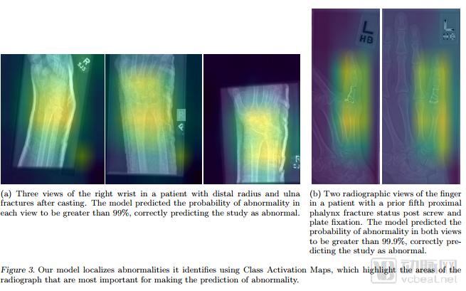
Models trained on MURA can accurately localize abnormalities in the wrist and fingers.
However, radiologists can still diagnose abnormalities in the elbow, forearm, hand, upper arm, and shoulder more accurately than the model.
![}P}E68_Q]3{~UWY9K4X6DZE.png](https://cdn.vcbeat.top/upload/image/07/01/19/21/1516346461561124.png)
△In the figure, green text indicates the best performance, while red text represents the worst. The model’s diagnostic performance for fingers and wrists surpassed that of the three radiologists. However, its diagnostic results for elbows and forearms were inferior to those of human experts.
This neural network comprises 169 layers. When multi-view X-rays of the upper extremities are input, the model can predict the probability of abnormalities.
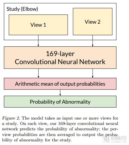
Workflow for Model Prediction of X-ray Abnormalities
As the largest medical imaging dataset, MURA contains 40,000 images, each individually annotated by radiologists.
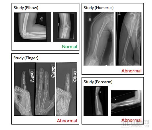
40,000 manually annotated X-ray images. Top left: normal elbow; top right: fractured upper arm; bottom left: degenerative changes in the distal phalanges; bottom right: ulna and radius fixed with plates and screws.
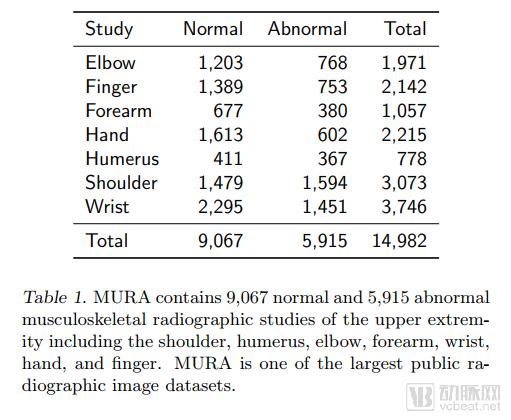
These 40,000 images are derived from nearly 15,000 research studies, of which 9,067 involve normal upper-limb skeletal muscle radiographs and 5,915 involve abnormal findings. The upper limb includes the shoulder, upper arm, elbow, forearm, wrist, hand, and fingertips.
Nowadays, an increasing number of datasets are emerging, providing excellent conditions for deep learning. The ability of AI algorithms to gradually surpass human performance in image recognition is largely attributable to the open-source availability of these datasets. Listed below are some currently available medical imaging datasets for your reference.
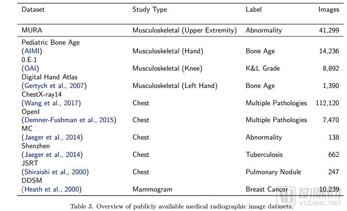
MURA is currently the largest open-source medical radiographic image dataset. The second-largest dataset, Pediatric Bone, contains 14,236 images and can be used to estimate bone age from hand X-rays. OAI is a knee joint dataset that can be used to detect knee osteoarthritis.
“However, given the current pace of AI development, medical schools should stop training students in radiology,” said Geoffrey Hinton, a professor in the Department of Computer Science at the University of Toronto, in an interview with The New Yorker.
The dataset will not be released until February; please continue to follow Stanford ML.
https://stanfordmlgroup.github.io/projects/mura/