Why Ultrasound + AI Has Become the Next Big Thing: A Multi-Billion-Dollar Opportunity Attracting Global AI Players
In 2017, the integration of medical imaging and artificial intelligence became a focal point of industry development, particularly in the combination of AI with radiological imaging. Both large corporations and startups achieved significant breakthroughs, with many products already undergoing clinical trials.
In fact, beyond radiological imaging, the application of artificial intelligence in the field of ultrasound has also garnered significant attention from the industry.
VCBeat (WeChat ID: vcbeat) has learned that global annual spending on ultrasound medical equipment exceeds $6 billion. Ultrasound imaging is widely adopted due to its advantages, including non-invasiveness, real-time image acquisition, and the absence of known side effects. However, the uneven quality of ultrasound diagnostic techniques and the shortage of skilled physicians at primary care levels have hindered the widespread adoption and application of ultrasound devices.
With breakthroughs in artificial intelligence technology, AI-assisted ultrasound diagnostic systems will help improve the accuracy of ultrasound diagnosis and accelerate the development of ultrasound equipment.
However, achieving AI-based diagnosis for ultrasound scan images requires significantly more research and is far more complex to process than simple medical image recognition. This is because ultrasound imaging possesses dynamic characteristics; when applying deep learning methods to make intelligent medical assessments from a dataset, it essentially involves analyzing ultrasound features from video sequences.。
Why is ultrasound-assisted diagnosis gaining attention? What are its underlying principles? Who are the key players in the market? And what are the major challenges to its development?
Principles of Ultrasound Diagnosis
Medical ultrasound examination, as a routine diagnostic modality, has been widely applied in clinical practice. Its working principle bears certain similarities to sonar: ultrasonic waves are emitted into the human body, where they undergo reflection and refraction upon encountering interfaces, and may be absorbed and attenuated within human tissues.
Because the morphology and structure of various human tissues differ, their reflection, refraction, and absorption of ultrasound waves also vary. Physicians identify these tissues based on the waveform patterns, curves, or imaging characteristics displayed by the diagnostic equipment.
The most widely used ultrasound diagnostic systems in clinical practice are B-mode and D-mode ultrasound, commonly referred to as B-scan ultrasonography and Doppler ultrasonography. Due to its advantages of being non-invasive, highly sensitive, broadly applicable, cost-effective, and easy to operate, ultrasound is extensively utilized in clinical diagnosis, particularly for the thoracic organs, heart, ophthalmology, and obstetrics and gynecology.
Two Reasons Why Ultrasound + AI Is Gaining Attention
Currently, with the development of internet technology, telemedicine has become a future trend due to its advantages such as speed and convenience. Although telemedicine certainly has its benefits, given the reliance of ultrasound on physicians' individual operational skills, current telemedicine operating models struggle to meet clinical work requirements.
Compared with the results of magnetic resonance imaging (MRI), computed tomography (CT), and electrocardiography (ECG), ultrasound diagnosis relies primarily on dynamic images acquired from various scanning planes by the operator, placing high demands on the sonographer’s technical proficiency. For the same lesion, variations in operators’ scanning techniques, image planes, instrument settings, and clinical experience may lead to differing diagnostic outcomes.
Telemedicine has largely addressed the issue of uneven distribution of high-quality medical resources. However, due to the specific nature of ultrasound medicine, large-scale clinical application of telemedicine in ultrasound diagnosis is not yet feasible at this stage, pending the establishment of standardized protocols for ultrasound image acquisition, image quality control, and transmission.
Therefore, leveraging auxiliary diagnostic systems to assist primary care physicians in addressing real-time diagnostic challenges may serve as a viable approach to mitigating the shortage of medical specialists at the grassroots level.
According to IHS statistics, the global market size for medical ultrasound diagnostic equipment reached approximately USD 6.2 billion in 2014 and is projected to reach USD 7.4 billion by 2019, with a compound annual growth rate (CAGR) of 3.6%.
In 2014, the market for ultrasound diagnostic equipment in China reached RMB 6.9 billion. Driven by the release of rigid demand, product upgrades and replacements, and the continuous advancement of favorable policies, the Chinese ultrasound equipment market is poised for rapid growth. The market size for medical ultrasound diagnostic equipment in China is projected to reach RMB 9.1 billion by 2019, with a compound annual growth rate (CAGR) of 5.7%.
In recent years, growing public attention to health has driven rapid development in the medical checkup industry. Compared with radiological examinations such as CT and MRI, non-invasive, radiation-free ultrasound examination is the preferred initial screening option for healthy or sub-healthy individuals. This trend will inevitably lead to continuous growth in the ultrasound equipment market and the number of ultrasound diagnostic procedures, thereby increasing physicians’ workload. The emergence of computer-aided diagnosis systems will help alleviate this situation.
Identification Principle
In the face of dynamic ultrasound imaging, each company has developed its own R&D strategy based on machine learning.
Bay Labs’ Solution: The U.S. startup employs intelligent video analytics to conduct detailed analysis of ultrasound scan videos and create user-specific models, accounting for the unique characteristics of each ultrasound dataset. This approach significantly simplifies the entire workflow of ultrasound imaging, from image acquisition and editing to clinical assessment.
Samsung Medison’s flagship device, the RS80A, features the S-Detect function, which employs deep learning algorithms to analyze breast lesions, enabling faster and more efficient ultrasound diagnostic imaging.
S-Detect provides recommendations for differentiating between benign and malignant lesions in selected clinical images, based on lesion characteristics derived from big data analysis of breast examination cases (100 million training samples).
S-Detect offers three modes, allowing users to adjust the sensitivity and specificity of automatic detection according to their requirements. The aim is to assist physicians in making more accurate diagnoses, while optimizing workflow and reducing repetitive operations.
The RS80A Ultrasound Diagnostic System adopts the American College of Radiology’s BI-RADS assessment categories as the standard for the standardized analysis and classification of suspicious breast lesions.
Dr. Song Yongqing, Chief Development Engineer of the Samsung RS80A and Head of S-Detect Technology R&D, and Dr. Park Moon-ho stated:
S-Detect employs a convolutional neural network technology customized for breast imaging. Once the user selects a seed point, the software processes all imported images to enhance the recognition of lesions with varying shapes, ultimately achieving automatic classification of the lesions.
This image transformation involves several critical steps, such as shifting, resizing, and warping, and is subsequently incorporated into a custom layer of the deep neural network.。
Furthermore, convolutional neural networks have the capability to independently learn from large volumes of training data to achieve image classification, which represents their most significant advantage over traditional machine learning methods. Composed of numerous convolutional layers, CNNs can automatically extract meaningful features from input images at specific layers.
These image features are then integrated within deeper layers. A learnable classifier is added at the end of the convolutional neural network, eliminating the need for developers to design hand-crafted features; instead, the convolutional neural network automatically extracts optimal features from the data during the learning process for classification purposes. This is precisely why Samsung Medical Center has chosen convolutional neural networks for the identification of breast lesions.
Kong Dexing, Chief Scientist at Deshang Yunxing and Qiushi Distinguished Professor at Zhejiang University, stated that the Deshang Yunxing AI-assisted diagnostic system for differentiating benign and malignant thyroid nodules is divided intoTraining and Scoring。
The training principle is based on deep learning technology, utilizing over 20,000 labeled ultrasound image samples to train the computer.
Traditional machine learning relies on manually defined features, such as the regularity of nodule margins and echogenicity patterns, which incorporate physicians’ subjective criteria. In contrast, deep learning eliminates the need for such handcrafted feature definitions.
Annotated samples are categorized into two major classes: benign and malignant. The neural network separately analyzes and extracts features associated with each class based on the annotations, a process that achieves greater granularity than visual inspection by the human eye.
Upon completion of training, Deshang Yunxing packages the algorithm and network parameters into a software application, namely the scoring system. This system can be deployed online or configured as a standalone version running on conventional computers, thereby enabling hospitals to utilize it for assisted diagnosis.
For the diagnosis of thyroid nodules, the average accuracy rate among physicians at tertiary hospitals currently stands at 60%–70%, while that at primary care hospitals is even lower. In contrast, the Deshang Yunxing AI-assisted diagnostic system now achieves an accuracy rate of over 85%.
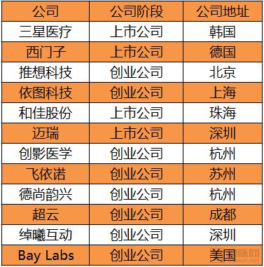
Over 10 Companies Deploying Ultrasound + AI
Unlike radiological imaging, not all medical AI companies have conducted research in the field of AI-powered ultrasound. According to incomplete statistics from VCBeat, companies involved in this research include Siemens, Samsung, Hejia Shares, Mindray, Infervision, Yitu Healthcare, Chuangying Medical, Vinno, Deshang Yunxing, Chaoyun, Shenzhen ChuoXi Interactive, and Bay Labs, among others.
These companies include traditional medical device manufacturers as well as innovative medical AI firms, all of which are actively entering this sector. For traditional device companies, leveraging AI to penetrate the market and enhance competitiveness is a sound strategy in an era where medical devices are becoming increasingly intelligent and portable. According to VCBeat, many companies are still in the early stages of researching AI-enhanced ultrasound, with considerable ground yet to be covered before commercial products can be realized.
Two Remaining Challenges to Address
However, some innovative medical AI companies have not ventured into the ultrasound sector. VCBeat learned from industry insiders that they highly value the "ultrasound + AI" field, as it features high frequency, substantial demand, and policy support, making it a promising area. The absence of involvement in "ultrasound + AI" is partly due to manpower constraints at startups, and partly due to two inherent challenges associated with ultrasound itself.
Low Level of Standardization. The Department of Ultrasound, like the Department of Radiology, also suffers from a shortage of outstanding physicians.However, unlike radiology, the greatest challenge in ultrasound does not lie in image interpretation, but rather in the proper operation of B-mode ultrasound equipment to obtain the specific images desired by the physician.For instance, there is currently no standardization regarding the amount of force required for patients of different body types, the specific hand gestures to be used, or how breathing should be adjusted; in practice, techniques often vary even among masters and their apprentices.
In contrast, CT or X-ray images are far more standardized than ultrasound images.
Workflow Issues。In China, sonographers interpret images and draft diagnostic reports simultaneously (with the exception of cardiovascular ultrasound), delivering results in near real-time. In contrast, radiologists first store the images and then review them at a later time. Consequently, ultrasound AI products face stringent requirements, necessitating chip-level integration with ultrasound equipment to enable real-time result generation.。
After all, it is difficult for doctors to lengthen their workflow for the sake of a good auxiliary software, which is a problem that developers need to think about.