South Korean AI Medical Imaging Leader Lunit Files for IPO After Outperforming IBM and Microsoft in Global Competitions

Lunit
Artificial Intelligence Software Developer
As research into artificial intelligence (AI) technologies deepens, their applications are increasingly extending across various industries. In recent years, computer-based image recognition accuracy has improved significantly. Applying AI technology to the field of medical image recognition offers substantial convenience to people. CIdi, a South Korean enterprise, is a startup founded on this innovative concept.
CIdi was founded in Seoul in August 2013. In October 2015, Cldi was renamedLunitabbreviated as Learning Unit, its primary area of expertise lies in processing medical imaging data. By leveraging a web-based medical image diagnostic software powered by cutting-edge deep learning technologies, it analyzes and interprets medical imaging data. This process extends beyond mere diagnosis, providing substantial support to physicians in making accurate, rapid, and effective clinical decisions.
Anthony is the CEO and co-founder of Lunit. He earned his Ph.D. in Electrical Engineering from the Korea Advanced Institute of Science and Technology (KAIST) in 2014. During his graduate studies, Anthony established a student-led deep learning group, whose members became the founding team of Lunit. As a multidisciplinary researcher, Anthony holds internationally recognized professional certifications in fields such as deep learning-based image recognition and semiconductor design.
Data-Driven Imaging Biomarker Technology (DIB Algorithm)
VCBeat has learned that, as an AI medical image analysis company, Lunit helps physicians make more accurate and effective clinical decisions through its Data-driven Imaging Biomarker (DIB) algorithms.
At the 2016 annual meeting of the Radiological Society of North America (RSNA), held from November 27 to December 2, 2016, at McCormick Place in Chicago, Illinois, Lunit debuted its data-driven imaging biomarker technology.

Data-Driven Imaging Biomarker (DIB) technology is an artificial intelligence-powered visual perception technology that is enabled by imaging biomarkers derived from large-scale medical image data.
DIB technology enables computers to accurately interpret medical images. By integrating large-scale medical imaging databases with deep learning techniques, it constructs advanced algorithms for automated detection and diagnosis, allowing users to rapidly scan medical images and obtain accurate diagnostic results.
DIB technology derives imaging biomarkers from large-scale medical image data, establishing a partnership between physicians and deep learning technologies. Traditional computer-aided detection (CAD) relies heavily on radiologists’ guidance, whereas DIB technology leverages the feature learning capabilities of deep convolutional neural networks to enable software systems to automatically identify significant diagnostic features from large-scale datasets.
Lunit's Three Major Products
Through continuous exploration, Lunit has applied its unique BID technology to medical imaging processing models, achieving high-accuracy recognition of chest X-rays, mammograms, and breast histopathology slides.

Chest radiography, first developed in 1900, is the most commonly used diagnostic imaging technique for evaluating pulmonary and cardiac abnormalities, aiding physicians in diagnosing various diseases (such as pneumonia, tuberculosis, and lung cancer) and monitoring treatment responses. Although chest radiography is a long-established technology, it continues to provide valuable reference information for clinical decision-making.
However, accurately interpreting chest X-rays remains a challenging task. It cannot be overlooked that, due to the inherent limitations of imaging equipment and the constraints of the human visual system, a considerable number of chest X-rays are misinterpreted by physicians, leading to misdiagnosis and subsequent worsening of patients’ conditions. Therefore, there is still room for improvement in the diagnostic accuracy and consistency of chest X-ray interpretation.
Lunit’s chest X-ray imaging leverages BID technology to gain deeper insights into pathological findings reflected in chest radiographs, enabling the development of enhanced computational models for lesion detection. These models are continuously trained via deep learning to improve the overall diagnostic performance of chest X-rays.
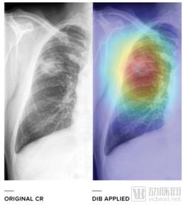
Comparison Before and After Using the BID Algorithm (Chest Radiography)
The application of DIB technology as a “second reader” in chest radiography demonstrates high diagnostic performance in detecting and differentiating abnormal lesions visible on chest X-rays, such as pneumonia, emphysema, diffuse lung diseases, pulmonary tuberculosis, lung cancer, and pulmonary metastases. DIB is expected to assist general practitioners and radiologists in interpreting chest X-rays more effectively and accurately.。
Mammography, first introduced in 1965, is the primary imaging modality for screening early-stage breast cancer. It employs low-dose radiation (approximately 0.7 mGy) to examine human breasts, predominantly in women, enabling the detection of various lesions such as breast tumors and cysts. This facilitates early diagnosis of breast cancer and reduces mortality rates. Mammography is widely utilized in national cancer screening programs worldwide. It plays a crucial role in earlier breast cancer detection, assessing the stage of the disease, and monitoring treatment response.
Mammography uses ionizing radiation to penetrate the human body for imaging, with radiologists analyzing the images for abnormalities. Despite continuous improvements, mammography has a false-negative rate (failure to detect existing cancer) of at least 10%, and a small number of patients experience false positives (abnormalities that are not cancer). The resulting high misdiagnosis rate has drawn considerable criticism within the medical community. Currently, mammography is far from perfect in terms of accuracy; given its widespread use worldwide, even modest improvements would benefit a large population of women.
Lunit introduces advanced BID algorithms to conduct detailed analysis of mammography images and has developed an improved malignant feature detection model, significantly reducing false-negative and false-positive misdiagnoses.。
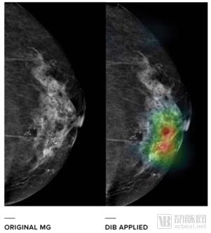
Comparison Before and After Using the BID Algorithm (Mammography)
The purpose of DIB in mammography is to improve the detection rate of breast cancer and reduce the misdiagnosis rate: 1) detect and localize lesions; 2) provide recommendations for the patient’s Breast Imaging-Reporting and Data System (BI-RADS); 3) predict the probability of lesion malignancy.。
Chest radiography and mammography are used for the initial detection and screening of diseases, while histopathological grading of breast tissue is a critical step in reaching a definitive medical diagnosis. Although pathological grading plays a vital role in the diagnostic process, the field still lacks quantifiable objective standards and detailed interpretive frameworks. The emergence of digital pathology offers hope for addressing these challenges.
Lunit has invested significant financial and human resources in digital pathology research to objectively interpret diverse morphological features in tissue samples, driving innovations that enhance the accuracy, efficiency, and consistency of histopathological diagnosis. Lunit is dedicated to developing state-of-the-art AI-based diagnostic software that automatically detects breast cancer in histopathology slides, classifies metastasis/spread, and assesses staging, thereby enabling pathologists to make more accurate and timely decisions.
In 2017, Lunit introduced an artificial intelligence algorithm capable of automated detection and staging assessment of breast cancer metastases in lymph nodes, marking the first attempt to fully automate a specific pathology task from end to end.。
When cancer is first diagnosed, the initial and most critical step is cancer staging. The international TNM staging system is a clinical staging method established by the Union for International Cancer Control (UICC). The TNM system classifies cancer pathologically, denoted as pTNM. Pathological diagnosis of the primary tumor (pT stage) requires resection of the primary tumor or biopsy procedures that allow for the most comprehensive assessment of the primary tumor tissue.
For the pathological diagnosis of regional lymph nodes (pN stage), a sufficient number of lymph nodes must be dissected to confirm regional lymph node metastasis. For the pathological diagnosis of distant metastasis (pM stage), histological examination is required. Based on the TNM staging system, the clinical stage of cancer patients can be clearly determined, prognosis assessed, and physicians guided in formulating the optimal treatment plan for each patient.
Pathological diagnosis of regional lymph nodes (pN staging: i.e., determining whether breast cancer has metastasized to the lymph nodes) involves processing an extremely large volume of image data, with images reaching a maximum resolution of 200,000 × 100,000 pixels. This requires pathologists to devote substantial time to carefully reviewing multiple images in order to accurately determine the pN stage.
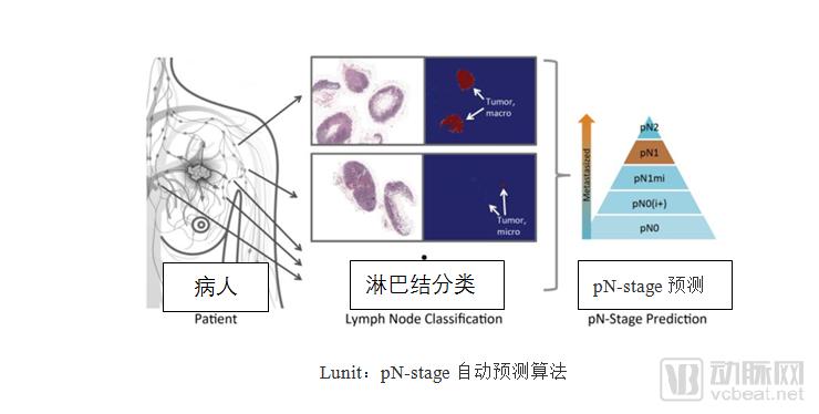
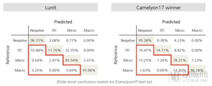
Lunit has leveraged its deep learning technology to develop a highly accurate pN-stage prediction algorithm. This algorithm integrates the detection and classification of tumor metastases across multiple lymph node tissue sections into a single clinical outcome. By utilizing lymph node histology images from the Camelyon17 dataset, Lunit established an algorithm for predicting pN-stage that outperforms most current state-of-the-art technologies worldwide, with the potential to significantly enhance pathologists’ efficiency and diagnostic accuracy.。
Lunit’s New AI-Powered Real-Time Imaging Platform—Lunit INSIGHT
Lunit returned to RSNA in 2017, bringing its latest cloud-based artificial intelligence real-time image analysis platform—Lunit INSIGHT—for its first public live demonstration.
Lunit’s real-time imaging platform has been extensively trained using deep learning algorithms. The 18 hospitals collaborating with Lunit have provided a vast dataset of de-identified clinical images, comprising over one million high-quality cases. Leveraging this image data, the system is trained to detect target diseases and radiological abnormalities, including lung cancer, tuberculosis, pneumonia, pneumothorax, and breast cancer in chest X-rays and mammograms.
Lunit INSIGHT is currently available to the public at https://insight.lun.io/. Users can upload their medical diagnostic images and receive AI analysis results from the software within seconds. Additionally, common features such as contrast adjustment and image zooming in/out are available within the software’s PACS viewer. The software has currently been integrated into the systems of multiple companies, including Nuance, EnvoyAI, and Infinitt Healthcare, for feasibility validation.
The promotion of technologies in the medical field must be supported by clinical validation. Suh, Chief Medical Officer at Lunit, stated, “Large-scale, multicenter studies on Lunit’s chest X-ray and mammography solutions will be conducted in early 2018, with validation results expected to be announced by late 2018. We also aim to obtain FDA approval for our chest X-ray and mammography solutions by the end of 2018.”
Clinical testing of chest X-ray images using the platform’s AI models—primarily for thoracic deformities, pulmonary nodules, lung masses, and pneumothorax—has demonstrated unprecedented accuracy, with standalone nodule detection reaching 97% and standalone detection of emphysema and pneumothorax achieving 99%.
According to the National Lung Screening Trial (NLST), the largest clinical trial conducted for lung cancer screening, results showed that chest X-rays missed 26.5% of lung cancer cases. Worldwide, over one billion chest X-ray examinations are performed annually; even a modest 10% reduction in the cancer miss rate would translate into substantial clinical benefits. In addition to solutions for chest X-ray and mammography, Lunit is also developing solutions for digital breast tomosynthesis, chest CT, and coronary CT angiography.。
Anthony: “Lunit’s vision is to develop advanced medical data analysis and interpretation software that surpasses human capabilities. With the launch of Lunit INSIGHT, we aim to contribute to ushering in a new era of medical practice by helping healthcare professionals make more accurate, consistent, and effective clinical decisions for their patients.”
Three rounds of financing exceeding $5 million
Lunit, as one of the leading international medical artificial intelligence companies,Lunit’s technology distinguishes itself from that of similar companies by being fully adapted to weakly labeled data, challenging the limits of human understanding in pattern recognition.。
Lunit’s deep learning technology has secured a prominent position on the international stage, featuring in numerous authoritative rankings:
Listed in CB Insights’ “AI 100” list of startups in 2017;
In 2017, it ranked first in the Camelyon Challenge. The Camelyon International Challenge is an international competition for machine-based diagnosis of pathological slides, jointly initiated by the Diagnostic Image Analysis Group (DIAG) and the Institute of Pathology at Radboud University Medical Center in the Netherlands. The aim of the challenge is to leverage machine learning to assist pathologists in the automated detection and classification of cancer metastases in breast cancer lymph node sections, thereby reducing subjective diagnostic errors and alleviating pathologists from heavy workloads.
Lunit ranked 5th in the 2015 ImageNet competition (the world's largest and most prestigious image recognition contest);
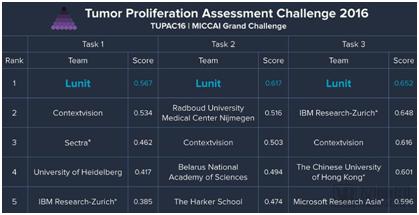
In the 2016 Tumor Proliferation Assessment Challenge (TUPAC), participants were tasked with predicting tumor proliferation scores under three different scenarios. Lunit secured first place in all three tasks, outperforming top companies such as Google, IBM (International Business Machines Corporation), and Microsoft. Since its establishment in 2013, the company has completed three rounds of financing, amounting to $3.51 million, $2 million, and $950,000, respectively, funded by leading venture capital firms Formation 8 and SoftBank Venture Capital.
Currently engaging in various large-scale research collaborations with KAIST and five major hospitals in South Korea., with its main industry competitors being Enlitic and Zebra Medical Vision.