Yizhan Medical and Tsinghua University Launch Breakthrough AI Solution for Carotid Plaque MRI Analysis
On April 26, the National Health Commission issued the “Notice on Further Strengthening the Management of Stroke Diagnosis and Treatment” (hereinafter referred to as the “Notice”). The Notice clearly stipulates the improvement of the national stroke diagnosis and treatment service system. It calls for vigorous promotion of the construction of emergency care systems, strengthening the provision of pre-hospital emergency equipment and facilities related to stroke diagnosis and treatment, and refining technical standards and operational procedures. The development of “Stroke Emergency Maps” is encouraged to establish “regional golden-hour treatment circles.”
Policy initiatives mark the growing national emphasis on stroke prevention. Stroke has become the leading cause of death in China, characterized by high incidence, high mortality, and high disability rates. Unstable vulnerable plaques in the internal carotid artery constitute an extremely high-risk factor for stroke; therefore, determining plaque integrity is a critical condition for preventing stroke occurrence.
The MRI Plaque Imaging AI Solution, jointly promoted by Wingspan Medical Group, the industrial translation platform of Tsinghua University’s Biomedical Imaging Research Center, and Beijing Qingying Huakang Technology Co., Ltd.It is precisely by leveraging artificial intelligence technology to address the medical imaging recognition of atherosclerotic plaques.。
Core Technological Advantages of the AI Solution for MRI Plaque Imaging
AI Solution for MRI Plaque ImagingLeveraging Tsinghua University’s strong engineering foundation, this AI solution was developed and generated based on tens of thousands of standard cases annotated by experts. The model provides accurate and comprehensive interpretation and analysis of plaque morphology and compositional characteristics, achieving a vessel wall segmentation accuracy of 97.2%. It assists physicians in rapidly locating lesions and assessing the condition of affected areas, thereby reducing workload, improving diagnostic efficiency, and enhancing diagnostic accuracy.
The system first analyzes image quality to determine usability, and provides feedback to physicians for rescanning of unusable images, thereby improving analytical accuracy.
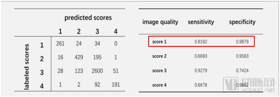
Evaluation Results for Sensitivity and Specificity on the Test Dataset (from the Tsinghua University Team)
Secondly, it automatically acquires multiple MR image sequences and performs image quality assessment, excluding substandard images while conducting image registration to generate multi-contrast vascular reconstruction images. It enables automatic identification of plaque structure and composition, completing precise carotid artery segmentation, plaque contour delineation, plaque component analysis, and detection of their three-dimensional distribution within minutes.
The diagnostic results employ the internationally recognized American Heart Association (AHA) standards for disease assessment and risk prediction, facilitating interventional prevention, treatment, and clinical decision-making by vascular surgeons, neurologists, interventional specialists, and cardiocerebrovascular physicians.
In the comparative experiments addressing rare cases, the contours manually delineated by radiologists were shown to be erroneous; in contrast, the model correctly segmented the carotid artery wall, fully demonstrating its robustness.
The left side of the figure below shows manual annotations, while the right side displays machine-generated annotations. It is evident that the machine has identified a fissure. The presence of a fissure indicates plaque rupture. Clinically, plaques with surface rupture are classified as high-risk plaques, as their contents can leak out, posing significant harm to patients and necessitating urgent diagnosis and treatment.
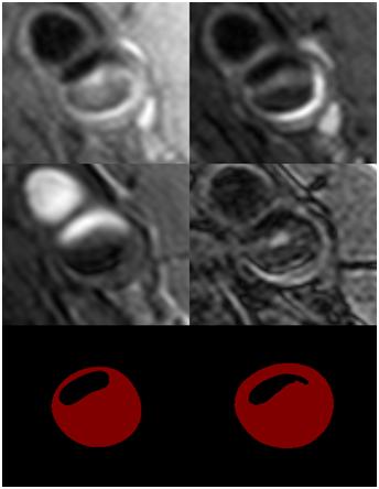
Left: manual annotation; Right: machine annotation (from the Tsinghua University team)
Unique Competitive Advantages of AI Solutions for Magnetic Resonance Plaque Imaging
The development of the magnetic resonance plaque imaging solution is based on the Tsinghua University Imaging Clinical Trial Project—Cardiovascular Risk Assessment in Chinese Population (CARE II). This project aims to utilize the latest magnetic resonance imaging techniques to screen, diagnose, provide risk预警, and monitor the prognosis of high-risk atherosclerotic plaques that cause ischemic stroke. It seeks to establish imaging diagnostic criteria suitable for the Chinese population, providing critical imaging evidence for the prevention and control of stroke among Chinese people.
During the experimental phase, the R&D team collaborated with 14 partner hospitals, including Beijing Hospital, Peking University First Hospital, and the 301 Hospital (PLA General Hospital), to collect over 30,000 sample data points from more than 1,000 patients across China. All samples were uniformly annotated by experts, creating the world’s largest standardized dataset for carotid artery plaques. Dozens of collaborating experts from top-tier tertiary hospitals in China, all holding titles of Associate Chief Physician or above and possessing over ten years of research experience in vascular plaque imaging, ensured the standardization and professionalism of the diagnostic content.

Dr. Li Rui
In addition to possessing the world’s largest standard dataset, the technical team behind the AI-powered MRI plaque imaging solution hails from the Biomedical Imaging Research Center at Tsinghua University. As pioneers who were among the first in China to explore this field, “it is no exaggeration to say that without this team, plaque imaging technology would not have emerged,” stated the product lead. The team members all graduated from top-tier universities abroad and bring years of theoretical expertise and hands-on R&D experience.
Dr. Li Rui, the lead of artificial intelligence algorithm development, is an Associate Researcher in the Department of Biomedical Engineering at the School of Medicine, Tsinghua University, and Deputy Director of the Center for Biomedical Imaging Research. His primary work focuses on the development of cardiovascular magnetic resonance imaging (MRI) techniques, with in-depth research particularly in intracranial and extracranial plaque imaging and blood flow imaging. He has served as principal investigator or co-investigator for two key projects under the 13th Five-Year Plan National Key R&D Program, three projects funded by the National Natural Science Foundation of China, one project under the Beijing Municipal Science and Technology Plan, one interdisciplinary project within Tsinghua University, and two industry-sponsored projects.
Clinical Application of AI Solutions for MRI Plaque Imaging
AI Solution for MRI Plaque Imaging connects to hospital Picture Archiving and Communication Systems (PACS) and leverages MRI plaque imaging data to assist physicians in performing precise analysis of arterial plaques across vascular beds, including the carotid and intracranial arteries, through qualitative and quantitative measurements. This solution reduces costs and enhances efficiency by freeing physicians from burdensome, repetitive tasks, allowing them more time for patient interaction and focus on higher-value activities, while providing a comprehensive solution for clinical diagnosis, treatment, and prognosis assessment.
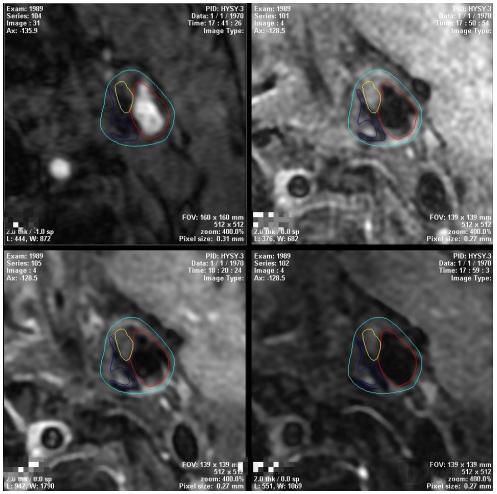
Typical Image of the Left Carotid Artery (from the Tsinghua University Team)
Currently, the clinical applications of AI-powered solutions for MRI-based plaque imaging primarily encompass three aspects. First, they assist physicians in characterizing plaque morphology, identifying intraplaque components, and differentiating between vulnerable and stable plaques, which holds significant predictive value for stroke occurrence. Second, they clarify the number and extent of plaque involvement, helping physicians define treatment objectives, guide therapeutic decisions, and assess prognosis. Finally, by automating report generation, these systems replace manual documentation, thereby enhancing diagnostic efficiency and accuracy, reducing patient wait times, and alleviating the burden on healthcare facilities.
The AI-powered solution for magnetic resonance plaque imaging boasts advanced technological advantages and immense application potential, delivering immeasurable value to clinical hospitals. This product meets the needs of hospitals, physicians, and the general public. By leveraging this intelligent tool, it enhances the quality of medical services for patients with cardiovascular diseases and elevates the standard of clinical care.
To better support hospitals and medical institutions, Wingspan Medical Group will announce its collaboration with the Biomedical Imaging Research Center of Tsinghua University at the “2018 Forum on Smart Medical Imaging” on July 1, 2018. By leveraging technological innovation, international cooperation, and industry integration, the partnership will combine Tsinghua University’s resources in medical imaging with Wingspan’s strong channel advantages to create a multi-modal, multidisciplinary research and clinical service platform.