Oral Digitalization as a Catalyst for Industry Upgrading: Strategic Layouts of Dentsply Sirona, Danaher, and Align Technology

Meyer
Developer of Core Technologies and Products for Intelligent Recognition
Digitalization is advancing into the core domains of dentistry. Early applications of digital technology in clinical dentistry primarily involved chairside fabrication of simple restorations and the use of surgical guides for dental implants. In recent years, digital technologies have been extensively adopted in prosthodontics, orthodontics, and implantology.
When employing fully digital jaw reconstruction, fully digital implantology, and digital orthodontics, software is used to execute various complex traditional procedural workflows. This simplifies and enhances the precision of intricate, time-consuming steps, allowing clinicians to visualize the treatment outcome before commencing any clinical procedures. This constitutes the essence of digital technology.
Reviewing the history of dental technology over the past few decades, we observe that many of the world’s most advanced technologies were first applied in the field of dentistry, such as lasers, 3D printing, CAD/CAM, implantology, three-dimensional design, and biomaterials. By integrating digital technologies into the artisanal craftsmanship of dentistry and transforming traditional craft-based processes into digital workflows, dental technology will evolve into a new era of high-tech digital solutions.
The digitalization of the dental industry, on the clinical side, generally refers to the adoption of CAD/CAM technology, intraoral scanners, and CBCT (Cone Beam Computed Tomography) to provide patients with more comprehensive treatment plans and an optimized care process, ultimately delivering better therapeutic outcomes, particularly in orthodontics and dental implantology. VCBeat (WeChat ID: vcbeat) attempts to outline and introduce the two foundational pillars of dental digitalization—intraoral scanning and CBCT—along with representative companies, highlighting the conveniences they bring to clinics, dentists, and patients.
Intraoral scanners have revolutionized the traditional clinical workflow for taking impressions and fabricating plaster models, eliminating numerous tedious steps such as waiting for impression materials to set, disinfecting impressions, and pouring models. This technology also reduces material and labor costs, enabling clinicians to directly acquire digital dental models and simplifying clinical procedures.
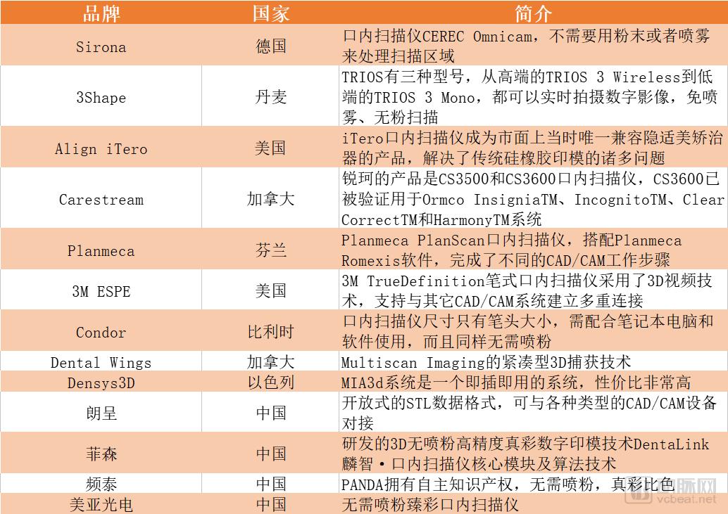
Mainstream Intraoral Scanners on the Market
Data acquired through intraoral scanning is processed by computer software to generate a comprehensive dataset containing all intraoral information required for both clinical and laboratory workflows. These data serve as the digital equivalent of physical impressions used in conventional restorative procedures. Once digitized, the digital impression files can be directly uploaded to the cloud, offering convenient storage, traceability, and reduced treatment duration for orthodontic cases.
By acquiring comprehensive intraoral data, a set of digital datasets can be integrated with other datasets, such as facial scans or 3D X-ray images. The results of big data analytics and simulation allow for multi-fold magnification of the intraoral condition, enabling more comprehensive and intuitive diagnosis and treatment planning.
These data can be leveraged for computer-aided diagnosis, treatment restoration, and planning, yielding more stable and effective outcomes than those based solely on physicians’ empirical experience. This represents the core clinical value of intraoral scanning. For instance, when integrated with software such as OraCheck and Cyfex, three-dimensional digital models enable visualized analysis of extensive intraoral changes—including tooth movement, tipping, rotation, gingival recession, and wear—thereby establishing a comprehensive digital orthodontic system.
In a study published not long ago in BMC Oral Health, the 3Shape TRIOS intraoral scanner was rated as the best among seven intraoral scanners tested, with the study comparing the clarity and accuracy of the abutment margin lines between traditional impressions and digital impressions.
For patients, they will enjoy efficient, comfortable, and intuitive precision treatment. Compared with traditional impression-taking, this approach is more comfortable, intuitive, safe, and precise, significantly reducing mouth-opening time and enhancing comfort. However, it comes at a relatively higher cost. Therefore, to some extent, digital products are more targeted toward mid-to-high-end customer segments.
Amid the workflow involving patients, dentists, clinics, and dental laboratories, dental laboratories are also facing the impact of digitalization. After dentists acquire digital impressions using intraoral scanners, they transmit the data via the cloud to dental laboratories for design. Once the laboratory experts complete the design, they immediately send the design files back to the clinic for milling and fabrication. While the patient remains at the clinic for treatment, the dental laboratory assumes the role of providing “design services.”
With the emergence of new restorative materials, dentists will be more inclined to opt for design services provided by dental laboratories. While dentists do not aspire to become technical experts themselves, they require chairside solutions to enhance the patient experience. For dental laboratories, this workflow hinges on a critical factor: digitalization.
Technicians at Glidewell Dental Lab, the largest dental laboratory in the United States, produce over one million digital dental restorations annually. The Glidewell.io™ digital solution, launched by the company in November 2017, was designed to shorten restoration delivery times and includes the iTero Element® intraoral scanner, fastdesign.io™ software, and the finalstage.io™ dental ceramic furnace.
In February 2018, Glidewell also partnered with Structo, a provider of dental 3D printing solutions, to integrate the Structo Velox desktop 3D printer into the glidewell.io™ In-Office solution. This integration enables dentists to access an all-in-one solution that includes intraoral scanning, chairside milling, and chairside 3D printing.
Currently, intraoral scanning systems remain expensive, with high-end models costing hundreds of thousands of yuan, which indirectly increases the operational costs for clinics and hospitals. However, their adoption is an inevitable trend for marketing purposes and enhancing profitability. Data indicates that 50% of dentists in the United States are considering purchasing intraoral scanners. Regardless of whether chairside systems are acquired, an increasing number of dentists will adopt intraoral scanning and transmit digital impression files. Intraoral scanners used by dental institutions in China are predominantly imported.
Foreign companies selling intraoral scanners in China must obtain a registration certificate and possess the relevant qualifications to sell their products through public channels. Domestic companies are also making strategic moves in this sector, such as Langcheng, Fussen, Pintech, and Hefei Meiya Optoelectronic Technology Inc. Users, including general hospitals and specialized dental hospitals, typically procure intraoral scanners through bidding processes.
As the regulatory framework for medical device registration continues to improve, it is expected that more intraoral scanner manufacturers will enter the Chinese market in the near future, leading to a decline in scanner prices.
3Shape
3Shape was founded in Copenhagen, Denmark, 15 years ago by Tais Clausen and Nikolaj Deichmann, who have played a pivotal role in driving the company’s rapid growth and establishing it as a global leader.
3Shape TRIOS intraoral scanners are used in clinics and laboratories across more than 100 countries. In 2005, the company launched the D200 scanner, marking its emergence in the dental field. In 2011, the 3Shape TRIOS intraoral scanner was officially released.
Currently, Trios is available in three models, ranging from the flagship Trios 3 Wireless to the more affordable Trios 3 Mono. This digital impression solution is widely adopted in the industry, enabling real-time capture of digital images with spray-free, powder-free scanning, and offering upgradable color modeling capabilities.
3Shape has launched a digital patient monitoring software tool that enables clinicians to compare intraoral scans over time. This software allows dentists to track and quantify gingival margin recession, tooth wear, and tooth movement. Digital solutions such as patient monitoring, orthodontic treatment simulators, patient dynamics, and digital shadow measurements are opening up new possibilities in dental care for both practitioners and patients.
Another critical aspect of digital technology is seamless integration. Regardless of whether a clinic has a chairside system, it can still utilize a wide variety of treatment protocols. 3Shape offers hundreds of orthodontic solutions, abutment libraries, and implant libraries—such as Straumann’s implant bridges and bars—and even includes ProSomnus’ sleep therapy solutions.
The advantage of having an open chairside solution is that it enables dentists to achieve true “single-visit treatment.” Even if a dentist has a chairside system but prefers not to perform any steps within their internal workflow, they can simply click to instantly share the file with a dental laboratory or solution provider, who will then complete the remaining work. Dentists with a digital mindset view soft tissues and available treatment options from a different perspective, opening up an entirely new world of possibilities.
In addition, the fully digital implant technology for edentulous patients in the United States has already applied the 3Shape Trios intraoral scanner to clinical scanning, while both European and South Korean fully digital implant technologies have adopted the 3Shape Trios intraoral scanning technology.
The key to fully digital techniques for implant-supported rehabilitation of edentulous jaws lies in the application of intraoral scanning. While various fully digital workflows differ, they all share the common feature of utilizing intraoral scans. Prior to implant placement, the Trios intraoral scanner can be used to scan both the maxillary and mandibular edentulous arches. The guide design software is then aligned with CBCT data, thereby eliminating errors associated with conventional impressions and stone models.
The fully digital workflow for implant-supported prosthetics in edentulous patients, as practiced in the United States, now enables preoperative design of bone reduction guides, surgical implant guides, immediate denture guides, and final restoration denture guides. This American approach represents a relatively mature strategy, wherein the final restorative prosthesis is milled from high-strength resin. A key feature is that all guides are designed preoperatively and used directly during surgery, eliminating waiting time for patients.
European fully digital technology for edentulous implantology now enables preoperative design of surgical guides. Immediate dentures and final prostheses are designed postoperatively by placing scan bodies intraorally and performing direct intraoral scanning. The immediate denture is fabricated using milling technology, while the final prosthesis can be completed using conventional digital workflows. South Korea’s fully digital technology for edentulous implantology is similar to the European approach.
China’s independently developed fully digital workflow for edentulous implant rehabilitation employs intraoral scanning of the edentulous arch, followed by 3D Digital Smile Design (DSD) for edentulous cases. The trial DSD prosthesis, radiographic prosthesis, bone reduction guide, surgical implant guide, immediate prosthesis guide, transitional prosthesis guide, and final restorative prosthesis guide are all designed using specialized software and fabricated via 3D printing or CAD/CAM milling. This approach enables a completely model-free restorative process and is currently considered a leading technology in the field.
In the Chinese market, the third-generation 3Shape TRIOS product has not yet been launched. Notably, on May 18, 3Shape and Angelalign signed a strategic cooperation agreement. Precise intraoral data acquisition is a critical prerequisite for the personalized customization of clear aligners. Through this partnership between Angelalign and 3Shape, users’ intraoral data can be rapidly and seamlessly integrated with Angelalign’s digital orthodontic platform, significantly driving innovation in digital dentistry.
Dentsply Sirona
Germany’s Sirona launched the CEREC system in 1987, marking an early entry into the field. Formerly known as Siemens Dental, Sirona is dedicated to developing and providing world-leading digital dentistry solutions, including CAD/CAM all-ceramic restoration systems for chairside (CEREC) and laboratory (inLab) use, dental treatment units, digital dental X-ray diagnostic systems, as well as dental instruments and sterilization equipment.
In March 2016, Sirona and Dentsply, the leader in the U.S. dental consumables market (Dentsply) became one of the world’s largest and most diversified manufacturers of professional dental products and technologies following the merger of equals.
In August 2012, Sirona’s newly launched intraoral scanner, CEREC Omnicam, was reviewed by a jury of 32 distinguished international experts. Recognized for its ergonomic design, it received the 2013 iF Product Design Award. The device supports video streaming, capturing moving images and reproducing the anatomical structures of the jaws in natural colors. Additionally, the camera features image stabilization.
The CEREC Omnicam intraoral scanner enables dentists to acquire digital impressions of intraoral teeth without the need for powder or spray treatment of the scanning area. During scanning, images of the patient’s oral cavity are displayed in natural color on the screen, allowing dentists to design high-precision all-ceramic restorations based on these highly accurate digital data.
Straumann
Straumann’s CARES comprehensive digital solution represents a complete dental workflow, from digital intraoral scanning to computerized production of restorations using state-of-the-art CAD/CAM processing. Each step seamlessly connects to the next, specifically designed for high-quality digital workflows.
This digital solution integrates intraoral scanning for digital data acquisition. If a clinic wishes to mill restorations in-house, it can use Straumann equipment to fabricate temporary crowns, inlays, onlays, and other restorations. Alternatively, if the clinic chooses to collaborate with a dental laboratory, Straumann provides laboratory workflows that leverage scanners to produce restorations. Additionally, Straumann offers CAD/CAM-based technologies; if chairside scanning is not feasible, clinics can send physical models or digital data to partner dental laboratories for fabrication.
CARES® Comprehensive Digital Solution is a workflow solution for exchanging and transmitting data among dentists, dental laboratories, and milling centers, offering an integrated end-to-end system. Straumann’s key differentiator lies in its open and selectable workflow, as opposed to a closed one.
iTero (Align Technology)
In 2011, Align (Align Technology) completed its acquisition of Cadent, making the iTero intraoral scanner the only product on the market at that time compatible with Invisalign aligners, thereby addressing many of the issues associated with traditional silicone rubber impressions.
Traditional PVS impressions are time-consuming, less comfortable, and cause a significant foreign-body sensation. The procedure typically takes about 30 minutes, and some patients may even experience nausea and vomiting. Subsequently, shipping the physical models to Align Technology in the United States requires approximately another two weeks. As a result, it often takes around one month to receive the final 3D treatment plan. Moreover, the prolonged shipping duration increases the risk of model deformation or damage, which can compromise accuracy and introduce errors.
The first step in the Invisalign treatment plan is taking impressions. The iTero intraoral scanner can complete a full-mouth digital scan within minutes, accurately capturing dental data. This process is not only fast but also allows for immediate generation of the ClinCheck treatment plan, providing a preliminary view of the orthodontic outcomes. The Invisalign 3D visualization plan can be viewed in as little as 48 hours, significantly reducing patient wait times and ensuring a comfortable overall experience.
A preliminary preview of the orthodontic outcome is available immediately after scanning, offering superior visualization that facilitates communication with dentists. As there is no need to ship physical models traditionally, dental data can be uploaded directly online, resulting in more accurate dental models.
The iTero digital impression system, since its launch in 2006 and after 12 years of development, has performed over 2.7 million orthodontic scans and officially entered the Chinese market in April 2018.
Dental imaging is currently undergoing a comprehensive transition toward digitalization, with cone-beam computed tomography (CBCT) being one of the key enabling technologies. In the 1990s, the first dental-specific CBCT unit, the NewTom 9000, was introduced, providing precise and clear three-dimensional facial images and enabling clinicians to obtain intuitive 3D surface representations of patients’ faces. The device entered the European market in 1996 and the U.S. market in 2001.
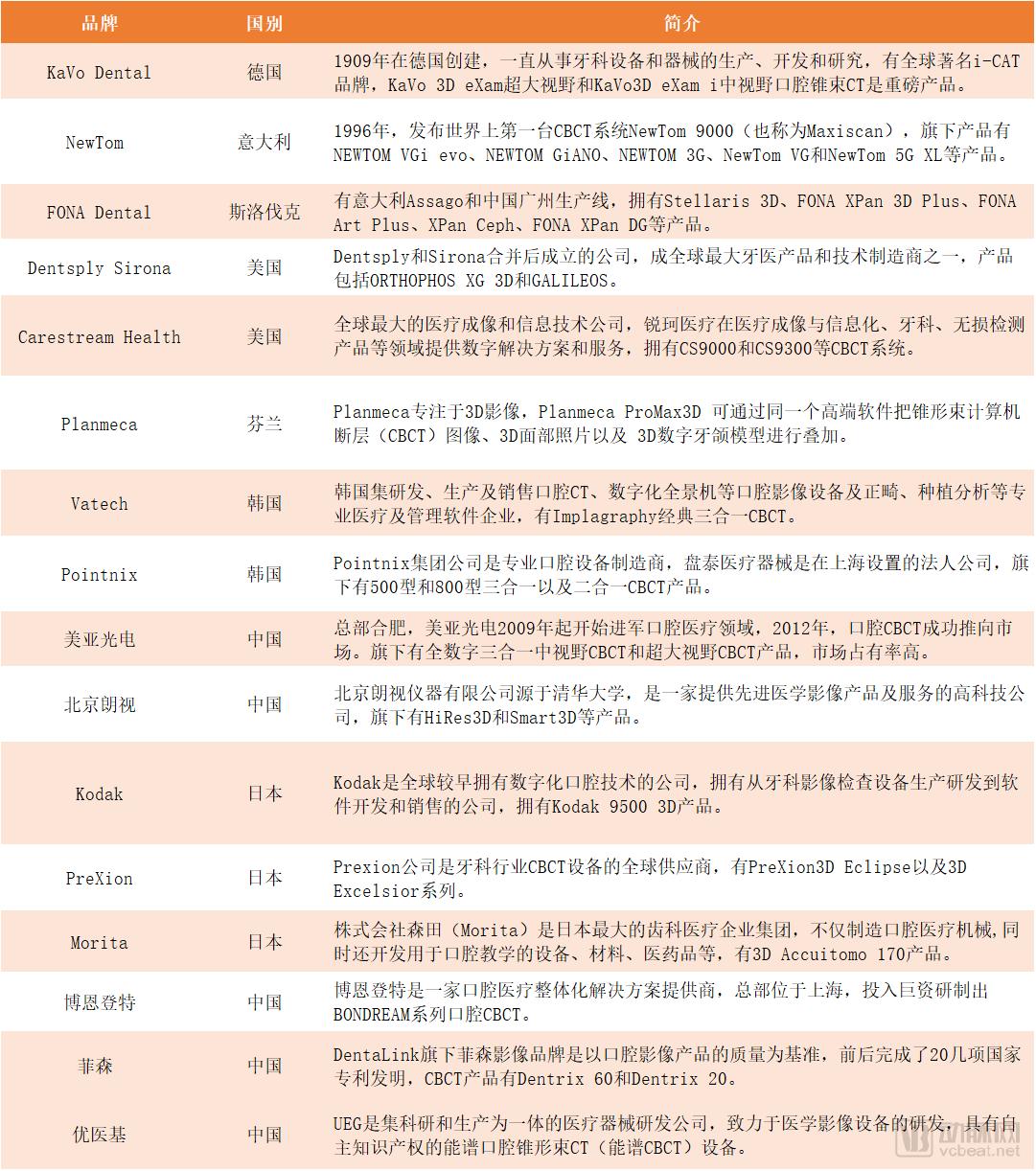
Mainstream CBCT Manufacturers and Products on the Market
Building upon traditional helical CT, cone-beam CT (CBCT) has upgraded the scanning and data acquisition methods. The most significant difference between the two is that CBCT employs a three-dimensional cone-beam X-ray scan to acquire nearly 600 distinct images, which are then reconstructed directly into a three-dimensional image with reduced metal artifacts. In contrast, traditional CT uses a two-dimensional fan-beam scan, generating two-dimensional image data after reconstruction, which is associated with more pronounced artifacts. CBCT is widely applied in fields such as oral and maxillofacial surgery, endodontics, orthodontic treatment, and dental implantology.
The advent of CBCT represents the evolution of medical imaging from two-dimensional to three-dimensional. CBCT has elevated scanning resolution to levels unattainable by spiral CT, even leading to the emergence of Micro-CT. Meanwhile, the radiation dose associated with CBCT has been reduced to a new low.
Without the application of CBCT in dentistry, digital orthodontics and dental implantology would not have been adopted as rapidly as they are today. CBCT has elevated scanning resolution to levels unattainable by spiral CT, even giving rise to Micro-CT. Meanwhile, the radiation dose associated with CBCT has been reduced to a new low.
For physicians, the matching software is relatively simple to operate, with fast and comprehensive 3D reconstruction capabilities; after training, even general technicians can operate it. For patients, the examination cost is correspondingly lower, making it more acceptable. For instance, when integrated with orthodontic software such as 3Shape and ExoCAD, CBCT enables physicians to pay closer attention to the thickness, height, and functional remodeling of the labial and lingual bone plates during orthodontic treatment. This is crucial for achieving high-quality orthodontic outcomes, long-term post-treatment stability, and periodontal health. Additionally, orthodontists are currently placing particular emphasis on the temporomandibular joint (TMJ), where CBCT plays a significant role in assessing condylar morphology and joint space.
Digital technology was first applied to dental implantology through the use of surgical guides, initially with guide designs generated by software developed by Simplant and Nobel. After more than a decade of clinical application and development, an increasing number of companies have introduced new guide design software, such as 3Shape, GuideMia, and ColorCube.
The principle of surgical guide design using implant planning software involves aligning CBCT data with dental models, simulating the position and orientation of implants via software, and determining their precise placement on the dental model. The software then generates a surgical guide, which can be fabricated through 3D printing or CAM milling. Most commercially available guide design software follows this workflow. For edentulous cases, radiographic dentures combined with CBCT data are commonly used for guide design.
The role of surgical implant guides is to facilitate fully or partially guided minimally invasive implant placement, as well as to determine the position and orientation of implants prior to flap elevation; in all cases, they are used to define and prepare the position and orientation of dental implants. Fully guided implant techniques have been applied in clinical practice for over a decade.
Fully Digital Implantology Technology, which originated in the United States in recent years, differs from fully guided implant surgery. Some mistakenly equate fully digital implantology with fully guided implant surgery. Fully digital implantology represents the comprehensive application of bone reduction guides, surgical implant guides, immediate denture guides, transitional denture guides, and final prosthetic restoration guides. In contrast, fully guided implant surgery relies solely on a surgical implant guide. Thus, fully digital implantology is a systematic strategy that integrates various guide-based technologies.
In dental implantology, prior to surgery, it is essential to assess the patient’s jawbone quality and volume, as well as to identify the locations of critical anatomical structures such as the inferior alveolar nerve canal and the maxillary sinuses. Based on this evaluation, the feasibility of the implant procedure is determined, along with key parameters including the type, dimensions, placement site, insertion trajectory, and depth of the dental implants.
With the assistance of implant planning software such as Simplant, NobelClinician, and 3Shape, CBCT devices can facilitate the design of implant surgeries and the fabrication of surgical guides, enabling precise control over implant position, angulation, length, and diameter. This ensures the success of both the surgical procedure and subsequent prosthetic restoration, making minimally invasive implant technology a recent trend.
Furthermore, its sensitivity, speed, and accuracy have been validated in the analysis of impacted teeth, as well as in the diagnosis of endodontic-periodontal diseases and temporomandibular joint disorders. Following immediate intraoral scanning, dentists can combine the data with CBCT imaging to design digital surgical guides for implants, enabling efficient and convenient preoperative planning. CBCT is an essential component of implantology services; any practice offering dental implants must be equipped with a CBCT system.
Currently, all reputable public hospitals are equipped with CBCT systems, primarily through centralized procurement, while spiral CT scanners are rarely used. Doctors, clinics, and hospitals have widely and fully accepted CBCT and its accompanying software. The three-dimensional perspective and tissue density analysis capabilities provided by CBCT enable physicians to obtain diagnostic results more conveniently and with greater confidence.
However, CBCT still holds a small market share, accounting for less than 5% when combining both public and private institutions. The key challenges to its adoption are, first, the cost of purchase, and second, training in software operation and imaging diagnostic techniques. In the future, spiral CT is unlikely to be used in teaching, diagnosis, or clinical practice, as it is a large-scale CT system requiring specialized personnel to operate and offers low resolution for soft tissues, which are the primary components in the oral cavity. Additionally, the accompanying dental diagnostic software needs further improvement.
In terms of pricing, large-field-of-view CBCT systems range from RMB 1.5 million to RMB 3.5 million, medium-field-of-view models from RMB 600,000 to RMB 1.5 million, and small-field-of-view units from RMB 400,000 to RMB 600,000. Small dental clinics are highly sensitive to upfront investment costs; if the initial outlay cannot be recouped within one to three years, their willingness to purchase is very low. Furthermore, dentists who have already acquired panoramic X-ray machines show little inclination to replace them with oral CBCT systems.
CBCT is an essential component of dental implant services; any facility offering implants must be equipped with a CBCT system. Introducing this new service line creates additional revenue streams for clinics and hospitals, making its adoption highly likely. However, purchasing decisions will ultimately depend on brand reputation, product quality, cost-effectiveness, and after-sales support. Additionally, there is significant market demand for supporting software solutions. Currently, available options are either imperfect or prohibitively expensive for high-quality systems. In the long run, software ecosystems will trend toward openness, inevitably leading to the emergence of a dominant platform.
The update cycle for CBCT systems depends on technological breakthroughs in key components, particularly X-ray detectors, averaging around five years, while accompanying software updates occur more frequently, typically every two years or so. Cost remains one of the primary barriers preventing smaller clinics from procuring digital equipment and software. Nevertheless, digital devices and technologies serve as marketing highlights and demonstrations of technical capability for clinics and hospitals, leading to strong willingness to invest in this area.
Carestream (Kodak)
Carestream provides medical and dental imaging systems, IT solutions, and advanced materials for the precision film and electronics markets worldwide. With operations in more than 150 countries and over 600 patented technologies in medical and dental imaging and information technology, Carestream has consistently been at the forefront of numerous technological advancements in medical imaging and healthcare IT.
Carestream Health, formerly known as the Eastman Kodak Health Group, carries forward Kodak’s legacy of innovation and has accumulated years of expertise in the field of medical imaging. The company holds more than 1,000 patents in medical and dental imaging and information technology.
The Rui CS Solution comprises a comprehensive suite of integrated systems and a range of open-system solutions, allowing physicians to flexibly select options that align with their clinical workflows and office layouts. Depending on the complexity of the case, clinicians can choose to perform scanning, design, and fabrication entirely within the clinic, or opt for in-office scanning only, transmitting digital impressions to a dental laboratory for further processing. This approach is highly convenient and efficient, significantly reducing patient wait times for treatment.
The latest product, CS 9300C Select, maintains a leading position in the CBCT market with four unique advantages: 3D CBCT imaging, digital CBCT impression scanning, panoramic imaging, and cephalometric imaging.
Danaher
Danaher Corporation (NYSE: DHR), founded in 1969 and formerly known as Diversified Mortgage Investors, Inc., adopted its current name in 1984. Headquartered in Washington, D.C., USA, the company is a global leader in the design and manufacture of science and technology innovation products and services. Its business is organized into four platforms: Life Sciences, Diagnostics, Dental, and Environmental & Applied Solutions.
A key milestone in Danaher’s development history was the acquisition of KaVo. Founded in 1909, KaVo is a German-headquartered leader specializing in the manufacture of dental equipment. Its core CBCT products include the i-CAT and KaVo 3D eXam.
In 2004, KaVo was acquired in its entirety by the Danaher Corporation, becoming a company under the Danaher Dental Products platform. In 2006, Sybron International Inc. of the United States was acquired by the Danaher Corporation, and KaVo Sybron became a leading global manufacturer of dental equipment and consumables, accounting for the largest share of Danaher’s dental business.
In 2017, Danaher’s dental business generated $2.819 billion in revenue, with the majority contributed by KaVo Kerr, ranking second globally in the dental sector, just behind the merged Dentsply Sirona.
This July, Danaher will spin off its dental business into an independent entity, DentalCo. The consolidation and development of DentalCo will enable greater operational efficiency, drive sustained margin expansion, place stronger emphasis on new investment opportunities, and support future earnings growth through strategic investments and mergers and acquisitions.
Meiya Optoelectronic
After more than six years of development, Meiya Optoelectronic has developed a full range of dental CBCT systems, including models with 12x8 cm medium field of view (FOV), 15x9 cm medium-large FOV, and 23x18 cm ultra-large FOV, fully meeting the needs of dental institutions at all levels. The company also offers complementary intraoral scanners, CAD, and CAM equipment, providing comprehensive, integrated solutions for patients' dental issues. It has established a product portfolio of dental X-ray CT diagnostic devices spanning medium, medium-large, and ultra-large FOVs, thereby satisfying customers' diverse product requirements.
Among them, the ultra-wide field-of-view CBCT offers an effective field of view as large as 23×18 cm in a single scan, meeting the clinical diagnostic and treatment needs of general dentistry and maxillofacial specialties. With over 2,000 customers in China and a market share exceeding 40%, it is a leading domestic CBCT brand.
Although digitalization is the trend, its adoption has not been as rapid as anticipated due to various factors, such as dentists’ established practices, high procurement costs for clinics, and the lengthy process for obtaining foreign medical device registration certificates. For instance, Invisalign’s iTero intraoral scanner officially entered the Chinese market in April this year, while 3Shape’s current offerings in China remain limited to its second-generation products.
Another major trend is that domestically produced equipment is increasingly matching international counterparts in performance while offering greater price advantages, accelerating the trend of domestic substitution. Currently, China has approximately 80,000 private dental clinics, which represent the primary growth driver for CBCT adoption.
References:
Jiang Shan: Fully Digital Design and Implant-Supported Prosthetic Technology for Edentulous Jaws
Today's Oral Health