Reaching the Top of "Radiology", Phased Array CT Rewrites the Evolution Path of Ultra-High-End

Nanovision
X-ray Detector and Static CT Product R&D Provider
In February 2026, the top international radiology journal Radiology published a highly anticipated prospective clinical study. The study focused on the clinical performance of using "dual-ring phased array static CT" for diagnosing small pulmonary nodules.
This study shows that the dual-ring phased array static CT independently developed by Nano-dimensional King (hereinafter referred to as "Nano-dimensional") performs excellently in the detection of tiny nodule lesions. With an ultra-fine slice thickness of 0.165mm and tens of millions of pixel-level imaging, the device is able to clearly capture pathological details of peripheral bronchi at levels 9 to 12 and tiny nodule lesions.

The realization of ultra-high-resolution imaging stems from the reconstruction of the imaging logic. The dual-ring phased array static CT transforms the principle of "mechanical rotation" imaging, instead seeking "optical rotation" imaging. It completes 360° data acquisition through sequential pulsed exposure from a large number of ray sources on the ring gantry, completely eliminating the centrifugal force limitations and artifact issues caused by mechanical rotation.

It is reported that the first phased array product "CompoundEye 24" of Beijing Nano-dimensional King Technology Co., Ltd. has recently obtained the Class III medical device registration certificate issued by the National Medical Products Administration (NMPA), securing the "pass" for commercial application.
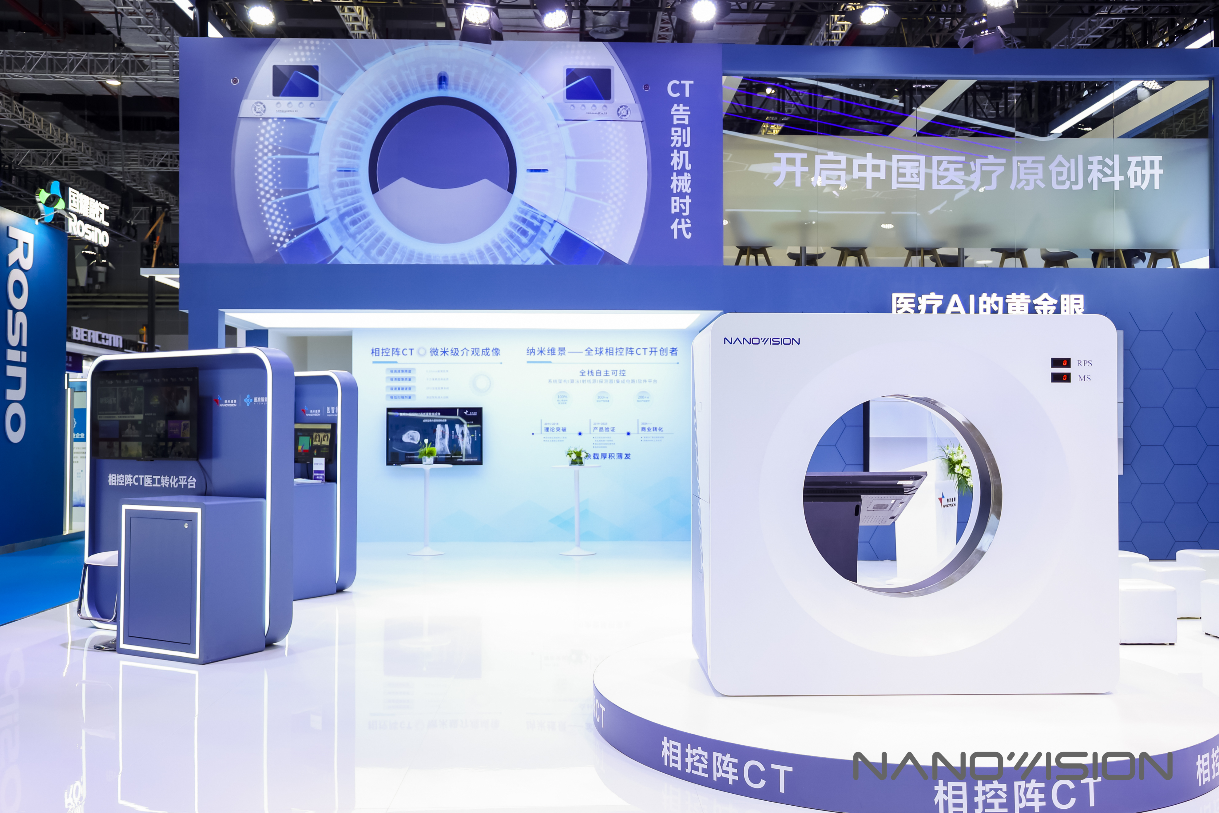
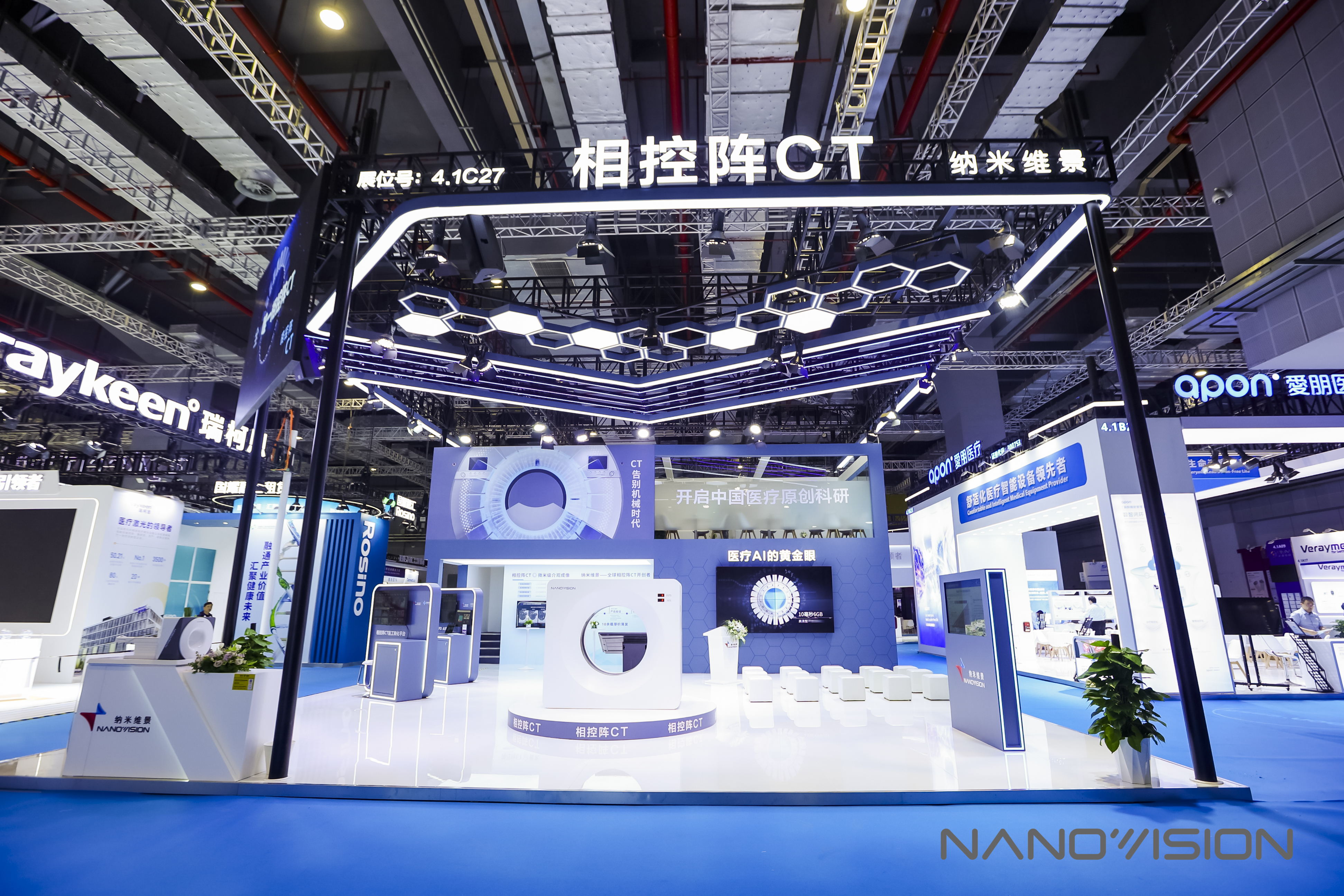
At the recently held 93rd China International Medical Equipment Fair (CMEF), phased array CT also made a strong entry into the imaging area with dual breakthroughs in top academic evidence and large-scale clinical approval, fiercely competing in the peak showdown of top CTs.
In the past 50 years, the development of spiral CT has always focused on the rotation speed of the tube-detector and the width of the detector.
But as the two elements approach their physical limits, CT manufacturers can no longer achieve a qualitative leap in image quality by iterating core components as they used to.
The physical limits brought by centrifugal force and the motion blur caused by high-speed rotation have become the "Achilles' heel" restricting the evolution of traditional spiral CT.
To break through the development bottleneck of spiral CT, Beijing Nano-dimensional King Technology Co., Ltd. has taken an alternative approach by creating "Phased Array CT". This innovation involves embedding a radiation source ring and a detector ring into the gantry of a single CT machine. Through precise electronic timing control, the radiation sources in the array are sequentially pulsed for exposure to complete data acquisition, replacing the mechanical rotation imaging of traditional CT.
Through "stationary source, moving light" electronic scanning, phased array CT avoids the tremendous centrifugal force generated by the high-speed rotation of spiral CT, completely eliminating centrifugal force and artifacts caused by mechanical rotation. Therefore, it can surpass the rotational speed limit of spiral CT, bringing about a qualitative change in spatial resolution.
Without the factor of "rotation speed," the technological iteration of phased-array CT will focus on the number of integrated tubes. As the number of tubes included in the ray source ring continues to increase, the capabilities of phased-array CT will further improve.
At this CMEF, Nano-dimensional King showcased its recently certified FlyEye 24, which features a ring of 24 integrated tube arrays and a detector ring with 64 detector module arrays. Relevant data indicates that this CT can achieve a reconstruction matrix of 3072×3072, with voxel accuracy reaching 0.165mm, and its spatial resolution far surpasses traditional spiral CTs.
In addition to achieving breakthroughs at the technical level, existing clinical trial evidence has also confirmed the value of phased array CT.
In the aforementioned study related to "Radiology," a comparative trial was conducted between phased-array CT and the world's top high-end spiral CT.
The results showed that: traditional CT can only image up to the 7th generation of bronchi, while phased array CT can display peripheral bronchi from the 9th to 12th generations; among 11 pulmonary structures, the visibility scores of 8 structures far exceeded those of traditional equipment.
It is important to note that the resolution breakthrough brought by phased array CT is not just for show; it indeed optimizes diagnostic decision-making by leveraging richer information.
For instance, in the diagnosis of ground-glass nodules (GGN), phased-array CT can accurately capture tiny lobulations on the nodule margins, internal vacuoles, and traversing blood vessels—these details are the core gold standard for distinguishing between benign and malignant nodules.
A lesion previously diagnosed as a "solid nodule" under traditional CT was accurately re-diagnosed as a "branch-like cavity structure" with the help of phased-array CT's micrometer-level perspective, thereby avoiding unnecessary surgical intervention.
Another study on phased array CT from the First Affiliated Hospital of Soochow University has also yielded encouraging results. The hospital's orthopedic team utilized high-definition, comprehensive, and low-radiation datasets generated by phased array CT to train a highly versatile "phased array CT + AI large model."
With the support of precise algorithms and a vast amount of clinical data, this model can significantly increase the early warning rate for osteoporotic fractures to 95%. It successfully addresses the industry pain points of high misdiagnosis rates and weak early warning capabilities in traditional diagnosis and treatment. This allows high-risk populations to be identified early and precisely intervened, truly achieving a leap from "precise diagnosis" to "proactive prevention."
Overall, the emergence of phased array CT not only represents an iteration in medical imaging technology but also redefines the future ecosystem of intelligent healthcare.
For medical AI, the tens of millions of pixels-level ultra-high-definition, structured data provided by phased array CT is the most ideal "fuel" for large models. It can not only improve the accuracy of automatic screening for early lung cancer, osteoporosis, and other diseases but also provide solid decision support for personalized treatment plans by mining radiomics information.
For clinical research, phased array CT is an unprecedented "observation tool." With an isotropic resolution of 0.165mm, it enables researchers to conduct studies at the "mesoscopic" level of the human body. Whether tracking subtle progressions in interstitial lung disease or analyzing mechanical changes in bone microstructure, more precise and comprehensive research data can be obtained, opening up entirely new avenues for medical research exploration.
In terms of humanized design, the 83cm ultra-large gantry aperture adopted by Nano-dimensional King far exceeds that of traditional CT equipment. This aperture not only better accommodates obese patients, alleviating the feeling of oppression and anxiety during examinations, but also promotes the integration of diagnosis and treatment, providing doctors with more operational space.
From the prototype in the laboratory, to the academic peak of "Radiology," to obtaining the NMPA registration certificate, and finally showcasing at the grand stage of CMEF, Beijing Nano-dimensional King Technology Co., Ltd. has spent a decade writing the story of China's high-end medical imaging resurgence.
Walking on the right path, Nano-dimensional King has the capability to bring China's high-end medical imaging technology to new heights, allowing more patients to benefit from the precise diagnosis and treatment offered by original technology. It also strengthens the voice of "Made in China" in the global medical field.